.
Brief Summary of the Guidelines for Diabetic Retinopathy
.
Landmarks in Diabetes
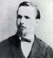
Paul Langerhans (1847-1888) 
discovered the Islets 
of Langerhans in 1869. 
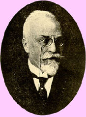
Oscar Minkowski (1858-1931)
discovered the development of
diabetes in pancreatomized dogs
in 1899.

Frederick Banting (1891-1941)
isolated insulin in 1921 
for human use.

BIBLIOGRAPHIC SOURCE(S)

  • American Academy of Ophthalmology Retina Panel, Preferred Practice Patterns Committee. Diabetic retinopathy. San Francisco (CA): American Academy of Ophthalmology (AAO); 2003. 33 p. [98 references]

BRIEF SUMMARY CONTENT

 RECOMMENDATIONS
 EVIDENCE SUPPORTING THE RECOMMENDATIONS
 IDENTIFYING INFORMATION AND AVAILABILITY


MAJOR RECOMMENDATIONS

The ratings of importance to the care process, (A, B, C) and the ratings for strength of evidence, (I, II, III) are defined at the end of the "Major Recommendations" field.

Diagnosis

The initial examination for a patient with diabetes mellitus includes all features of the comprehensive adult medical eye evaluation, with particular attention to those aspects relevant to diabetic retinopathy.

History

An initial history should consider the following elements:

  • Duration of diabetes [A:I]
  • Past glycemic control (hemoglobin A1c) [A:I]
  • Medications [A:III]
  • Medical history (e.g., onset of puberty, [A:III] obesity, [A:III] renal disease, [A:II] systemic hypertension, [A:I] serum lipid levels, [A:II] pregnancy [A:I])
Examination

The initial examination should include the following elements:

  • Best-corrected visual acuity [A:I]
  • Intraocular pressure [A:III]
  • Gonioscopy when indicated [A:III]
  • Slit-lamp biomicroscopy [A:III]
  • Dilated funduscopy including stereoscopic examination of the posterior pole [A:I]
  • Examination of the peripheral retina and vitreous [A:III]
Slit-lamp biomicroscopy with accessory lenses is the recommended method to evaluate retinopathy in the posterior pole and midperipheral retina. [A:III] The examination of the peripheral retina is best performed with indirect ophthalmoscopy or with slit-lamp biomicroscopy, combined with a contact lens. [A:III]

Examination Schedule

Recommended Eye Examination Schedule for Patients with Diabetes Mellitus
 
Diabetes Type Recommended Time of First Examination Recommended Follow-up*
Type 1 5 years after onset [A:II] Yearly [A:II]
Type 2 At time of diagnosis [A:II] Yearly [A:II]
Prior to pregnancy (type 1 or type 2) Prior to conception or early in the first trimester [A:I] No retinopathy to mild or moderate nonproliferative diabetic retinopathy (NPDR): every 3-12 months [A:I] 

Severe NPDR or worse: every 1-3 months [A:I]

*Abnormal findings may dictate more frequent follow-up examinations.

Treatment

Management recommendations for patients with diabetic retinopathy are summarized in the table below.

Management Recommendations for Patients with Diabetes
 
Severity of Retinopathy Presence of clinically significant macular edema (CSME1) Follow-up (Months) Scatter (Panretinal) Laser Fluorescein Angiography Focal Laser2
1. Normal or minimal NPDR No 12 No No No
2. Mild to moderate NPDR No 

Yes

6-12 

2-4

No 

No

No 

Usually

No 

Usually1, 3

3. Severe or very severe NPDR No 

Yes

2-4 

2-4

Sometimes4

Sometimes4

Rarely 

Usually

No 

Usually5

4. Non-high-risk PDR No 

Yes

2-4 

2-4

Sometimes4

Sometimes4

Rarely 

Usually

No 

Usually3

5. High-risk PDR No 

Yes

3-4 

3-4

Usually 

Usually

Rarely 

Usually

No 

Usually5

6. High-risk PDR not amenable to photocoagulation (e.g., media opacities) -- 1-6 Not Possible6 Occasionally Not Possible6

  1. Exceptions include: hypertension or fluid retention associated with heart failure, renal failure, pregnancy, or any other causes that may aggravate macular edema. Deferral of photocoagulation for a brief period of medical treatment may be considered in these cases. Also, deferral of CSME treatment is an option when the center of the macula is not involved, visual acuity is excellent, close follow-up is possible, and the patient understands the risks.
  2. Focal photocoagulation refers to direct focal laser to leaking microaneurysms or a grid photocoagulation pattern to areas of diffuse leakage or nonperfusion seen on fluorescein angiography.
  3. Deferring focal photocoagulation for CSME is an option when the center of the macula is not involved, visual acuity is excellent, close follow-up is possible, and the patient understands the risks. However, initiation of treatment with focal photocoagulation should also be considered because, although treatment with focal photocoagulation is less likely to improve the vision, it is more likely to stabilize the current visual acuity.
  4. Scatter (panretinal) photocoagulation surgery may be considered as patients approach high-risk PDR. The benefit of early scatter photocoagulation at the severe nonproliferative or worse stage of retinopathy is greater in patients with type 2 diabetes than in those with type 1. Treatment should be considered for patients with severe NPDR and type 2 diabetes. Other factors, such as poor compliance with follow-up, impending cataract extraction or pregnancy, and status of the fellow eye will help in determining the timing of the scatter photocoagulation.
  5. Some experts feel that it is preferable to perform focal photocoagulation first, prior to scatter photocoagulation, to minimize scatter laser-induced exacerbation of the macular edema.
  6. Vitrectomy is indicated in selected cases.
Follow-up

The follow-up evaluation includes a history and examination.

History

A follow-up history should include changes in the following:

  • Symptoms [A:III]
  • Systemic status (pregnancy, blood pressure, renal status) [A:III]
  • Glycemic status (hemoglobin A1c) [A:I]
Examination

A follow-up examination should include the following elements:

  • Visual acuity [A:I]
  • Intraocular pressure [A:III]
  • Slit-lamp biomicroscopy with iris examination [A:II]
  • Gonioscopy (if iris neovascularization is suspected or present or if intraocular pressure is increased) [A:II]
  • Stereo examination of the posterior pole with dilation of the pupils [A:I]
  • Peripheral retina and vitreous examination, when indicated [A:II]
Recommended intervals for follow-up are given in the above table.

Provider

Because of the complexities of the diagnosis and surgery for PDR, the ophthalmologist caring for patients with this condition should be familiar with the specific recommendations of the Diabetic Retinopathy Study (DRS), Early Treatment Diabetic Retinopathy Study (ETDRS), United Kingdom Prospective Diabetes Study (UKPDS), and Diabetes Control and Complications Trial (DCCT). [A:III] The ophthalmologist should also have training in and experience with the management of this particular condition. [A:III]

Counseling/Referral

Patient education about the importance of maintaining near-normal glucose levels and near-normal blood pressure and lowering serum lipid levels is an important aspect of the care process. [A:III]

Patients with diabetes mellitus without diabetic retinopathy should be encouraged to have annual dilated eye examinations to detect the onset of diabetic retinopathy. [A:III] Patients should also be informed that effective treatment for diabetic retinopathy depends on timely intervention, despite good vision and no ocular symptoms. [A:III]

Those patients whose conditions fail to respond to surgery and those for whom further treatment is unavailable should be provided with proper professional support and offered referral for counseling, vision rehabilitation, or social services as appropriate. [A:III]

Definitions:

Ratings of Importance to Care Process

Level A, most important
Level B, moderately important
Level C, relevant but not critical

Ratings of Strength of Evidence

  1. Level I includes evidence obtained from at least one properly conducted, well-designed randomized, controlled trial. It could include meta-analyses of randomized controlled trials.
  2. Level II includes evidence obtained from the following:
    • Well-designed controlled trials without randomization
    • Well-designed cohort or case-control analytic studies, preferably from more than one center
    • Multiple-time series with or without the intervention
  3. Level III includes evidence obtained from one of the following:
    • Descriptive studies
    • Case reports
    • Reports of expert committees/organization
    • Expert opinion (e.g., Preferred Practice Pattern panel consensus)

CLINICAL ALGORITHM(S)

None provided

EVIDENCE SUPPORTING THE RECOMMENDATIONS


TYPE OF EVIDENCE SUPPORTING THE RECOMMENDATIONS

The type of supporting evidence is identified and graded for each recommendation (see "Major Recommendations.")

IDENTIFYING INFORMATION AND AVAILABILITY

BIBLIOGRAPHIC SOURCE(S)
  • American Academy of Ophthalmology Retina Panel, Preferred Practice Patterns Committee. Diabetic retinopathy. San Francisco (CA): American Academy of Ophthalmology (AAO); 2003. 33 p. [98 references]

ADAPTATION

Not applicable: The guideline was not adapted from another source.

DATE RELEASED

1998 Sep (revised 2003)

GUIDELINE DEVELOPER(S)

American Academy of Ophthalmology - Medical Specialty Society

SOURCE(S) OF FUNDING

American Academy of Ophthalmology

GUIDELINE COMMITTEE

Preferred Practice Patterns Committee, Retina Panel

COMPOSITION OF GROUP THAT AUTHORED THE GUIDELINE

Retina Panel Members: Emily Y. Chew, MD (Chair); William E. Benson, MD; H. Culver Boldt, MD; Tom S. Chang, MD; Louis A. Lobes, Jr., MD; Joan W. Miller, MD; Timothy G. Murray, MD; Marco A. Zarbin, MD, PhD; Leslie Hyman, PhD (Methodologist)

Preferred Practice Patterns Committee Members: Joseph Caprioli, MD (Chair); J. Bronwyn Bateman, MD; Emily Y. Chew, MD; Douglas E. Gaasterland, MD; Sid Mandelbaum, MD; Samuel Masket, MD; Alice Y. Matoba, MD; Donald S. Fong, MD, MPH

FINANCIAL DISCLOSURES/CONFLICTS OF INTEREST

No proprietary interests were disclosed by members of the Preferred Practice Patterns Retina Panel for the past 3 years up to and including June 2003 for product, investment, or consulting services regarding the equipment, process, or products presented or competing equipment, process, or products presented.

GUIDELINE STATUS

This is the current release of the guideline.

This guideline updates a previous version: American Academy of Ophthalmology (AAO), Preferred Practice Patterns Committee, Retina Panel. Diabetic retinopathy. San Francisco (CA): American Academy of Ophthalmology (AAO); 1998. 32 p.

All Preferred Practice Patterns are reviewed by their parent panel annually or earlier if developments warrant.

GUIDELINE AVAILABILITY

Electronic copies: Available from the American Academy of Ophthalmology (AAO) Web site.

Print copies: Available from American Academy of Ophthalmology, P.O. Box 7424, San Francisco, CA 94120-7424; telephone, (415) 561-8540.

AVAILABILITY OF COMPANION DOCUMENTS

None available

PATIENT RESOURCES

The following patient education booklet is available:
  • Diabetic retinopathy (2001)
The following patient education brochure is available:
  • Diabetic retinopathy (1998)
  • Diabetic Retinopathy (Spanish: Retinopatia Diabetica) (1998).
The following patient education videotape is available:
  • Diabetic retinopathy (1990)
Print copies: Available from the American Academy of Ophthalmology (AAO), P.O. Box 7424, San Francisco, CA 94120-7424; Phone: (415) 561-8540.

Please note: This patient information is intended to provide health professionals with information to share with their patients to help them better understand their health and their diagnosed disorders. By providing access to this patient information, it is not the intention of NGC to provide specific medical advice for particular patients. Rather we urge patients and their representatives to review this material and then to consult with a licensed health professional for evaluation of treatment options suitable for them as well as for diagnosis and answers to their personal medical questions. This patient information has been derived and prepared from a guideline for health care professionals included on NGC by the authors or publishers of that original guideline. The patient information is not reviewed by NGC to establish whether or not it accurately reflects the original guideline's content.

NGC STATUS

This summary was completed by ECRI on February 20, 1999. The information was verified by the guideline developer on April 23, 1999. This summary was updated again on April 30, 2004. The information was verified by the guideline developer May 20, 2004.

COPYRIGHT STATEMENT

This NGC summary is based on the original guideline, which is subject to the guideline developer's copyright restrictions. Information about the content, ordering, and copyright permissions can be obtained by calling the American Academy of Ophthalmology at (415) 561-8500.
Epidemiology Clinical features Risk factors Screening
Lasers and lenses. NVD,, NVE.. Maculopathy
Vitrectomy. Cataract Special problems Counselling
References.. AAO guidelines Atlas of Retinopathy Contact lenses
Main index Main page.