General examination in open angle glaucoma
 

Cystic bleb indicating a functioning 
trabeculectomy

Adrenochrome pigments on the tarsal 
conjunctiva seen with topical adrenaline use

In the examination, the examiner may ask you to examine a patient with known open angle glaucoma.

You need to look for :

  • physical signs indicating the treatment that the patient is receiving
  • physical signs indicating the possible causes such as pigment dispersion syndrome and pseudoexfoliation syndrome
  • physical signs indicating the severity of the lesion
The most common sign is trabeculectomy. This is usually performed at the superior limbus. However, if there was previous 
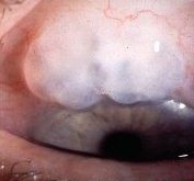
operation the second trabeculectomy may be found more temporally. Iridectomy is usually present. Poor drainage is 
suggested by an absence of bleb or a dome-shaped vascularized bleb caused by subconjunctival fibrosis. Occasionally, you
may encounter patients who had Scheie procedure for aqueous drainage. This full-thickness filtering surgery. The 
anterior chamber is opened to the subconjunctival space. Uveal tissue is visible at the sclerotomy site. This procedure 
is now out-dated.

Other physical signs associated with glaucoma treatment include:

  • constricted pupil with pilocarpine
  • heterochromia iridis with latanoprost especially if the application is unilateral
  • adrenochrome with topical adrenaline application, the conjunctiva is usually hyperaemic from the use of adrenaline.

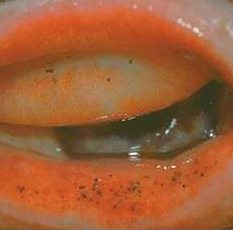
  • This can be easily missed unless you evert the lower lid which it is most commonly seen.
  • Molteno's tube in the anterior chamber


The most common cases of secondary open angle glaucoma with anterior physical signs are pigment dispersion syndrome
pseudoexfoliation syndrome, iridocorneal endothelial (ICE) syndrome and Fuch's heterochromic cyclitis.

The severity of the glaucoma is graded according to the loss of neural tissue best seen at the optic disc.
 

Questions:

1. What are the advantages and disadvantages between limbal based and fornix based conjunctival flap in trabeculectomy?

2. How would you manage a flat anterior chamber post-trabeculectomy?

3. What factors increase the risk of failure in trabeculectomy?


Return to the main page