Pigment dispersion syndrome
 

Radial transillumination of the iris in the
midperiphery region. This is seen with
retroillumination. Each transillumination
represents area devoid of pigment
epithelium

Krukenberg's spindle
with diffuse illumination

Krukenberg's spindle 
with retroillumination

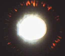
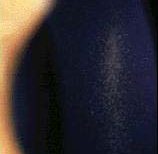
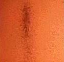
The corneal endothelium contains vertically orientated deposition of pigments (Krukenberg's spindle).
The pigment may also be seen on the iris and the lens (and also the trabecular meshwork but in the clinical 
examination you are unlikely to be asked to perform gonioscopy on the patient). Retroillumination shows 
mid-periphery iris transillumination.

In the examination:

  • look for any peripheral iridoctomies which may be performed in an attempt to reduce the production

  • of pigment
  • look at the patient's glasses, most of this patients has myopia
  • mention you like to examine the optic disc for cupping

Questions:

1. What proportion of patients with pigment dispersion syndrome develop glaucoma?

2. Is it possible for a patient with pigment dispersion syndrome to develop glaucomatous disc changes despite a normal intraocular pressure and if so why?


3. What is the cause of the Krukenberg's spindle?

4. What is the theoretical advantage of peripheral iridotomies in pigment dispersion syndrome?


Return to the main page


return to the top