Common anterior chamber cases
(click on the picture or condition for case descriptions and questions)

In the clinical examination, the anterior chamber should be examined for:
  • cells and flare 
  • foreign bodies (Molteno's tube and silicone oil and less commonly heavy liquid)
  • depth of the anterior chamber if you suspect the anterior chamber is

  • shallow especially if there were signs of previous acute angle closure glaucoma 
    (the angle is assessed by the van Herick's method and the depth of the 
    anterior chamber is measured using the method described in the section on 
    common examination techniques) 

Cells and flare

Molteno's tube

silicone oil 
Return to the main page


van Herick's technique
(depth of peripheral anterior chamber (PAC) compared to the adjacent 
 corneal thickness (CT) at temporal corneal limbus at 60° angle)

The technique uses the following set-up:

  • optical section
  • 60° between observation and illumination 
  • full slit length 
  • magnification approximately x 15 
  • low to medium illumination 
  • place optical section just inside limbus. 

  • Assuming the corneal thickness = 1 unit, assess the width of the 
    "aqueous gap" or peripheral anterior chamber from corneal 
    endothelium to iris
     
     
    Grade  Ratio of aqueous gap/cornea clinical interpretation
    4 > 1/2 : 1 closure impossible
    3 = 1/2 - 1/4 : 1 closure impossible
    2 = 1/4 : 1 closure possible
    1 < 1/4:1 closure likely with full dilatation
    0 nil closed

     
    Return to the top


Cells and flare in the anterior chamber
 

Mutton fat keratic precipitates

Fine stellate keratic precipitates in 
Fuch's heterochromic cyclitis

Koeppe nodules at the pupil margin

Busacca nodule
.
Cells and flare are common signs in uveitis. You need to look for other associated signs which may point to the type 
of inflammation. Most candidates should have not problems spotting mutton fat precipitates but stellate precipitates
may be easily missed if they were small and sparse.
The other signs which may be missed are granulomas on the iris. The Koeppe nodules are found at the pupil margin 
and tend to be smaller and more common than Busacca nodules. Both conditions are indicative of granulamatous 
inflammation.
 
 
Return to the top
.


Molteno's tube
 

Molteno's tube can usually be easily spotted. However, if the tube is short, it may not be apparent unless you 
get the patient to look down. 

Molteno's tube is usually associated with the following signs:

  • previous trabeculectomy (very often there are more than one trabeculectomy with several iridectomies) 
  • neovascular glaucoma 
  • iridocorneal endothelial syndrome
  • advanced glaucomatous disc.
Return to the top


Silicone oil and heavy liquid
 

Emulsified silicone oil in the anterior chamber
(hyperoleon)

Globules of heavy liquid in inferior anterior chamber
.
With the advance of vitreoretinal surgery, the above physical signs are becoming more common in the clinical examination. 
Always ask to examine the posterior segment for the presence of previous retinal detachment, proliferative vitreoretinopathy 
and advanced proliferative diabetic retinopathy. 

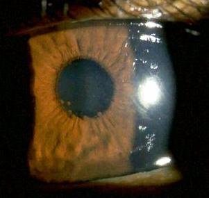
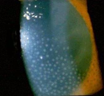
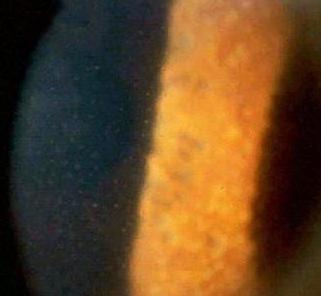
Silicone oil in the anterior chamber

The superior anterior chamber contains fine suspension of silicone oil. The oil may appear milky owing to emulsification
(so-called inverted hypopyon).
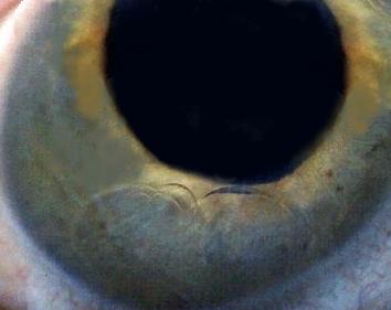
Look for:

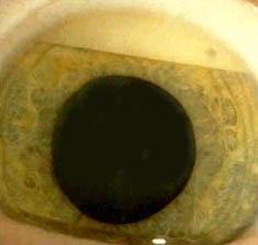
  • complications associated with the oil (ie. band keratopathy and cataract)
  • presence of Anton's iridotomy (this is iridectomy performed at 6 O'clock in aphakic patient to prevent 

  • pupillary block)
  • previous retinal detachment
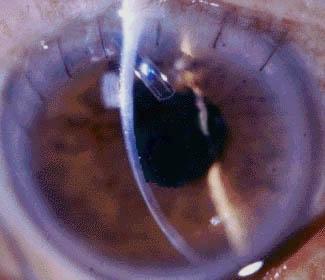
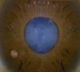
Heavy liquid in the anterior chamber

There are globules of liquid in the inferior part of the anterior chamber. 

Look for:

  • previous retinal detachment operation 
.

Questions:

1. What are the uses of silicone oil?

Return to the top