Eyelids and anterior segment: Case eleven
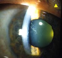
This 60 year-old woman was referred by her family doctor because of several episodes of sharp pain in her right eye over the past few months. Her visual acuity was normal in both eyes and the intraocular pressures were normal. The above picture is the appearance of her right anterior segment.a. What does the picture show?
Whitish anterior subcapsular opacities in a lacy pattern.This is a typical appearance of glaucoflecken (glaucoma flecks). Glaucuoflecken is caused by pressure necrosis of anterior lens epithelium with adjacent subcapsular cortical degeneration. The opacities tend to diminish gradually and become sparse. As new lens fibres grow in form the equator, they overlie the opacities which sink deeper into the lens but will persist as a permanent sign of pressure necrosis.
b. What other physical signs may be present?Further examination will show shallow anterior chamber and narrow angle of the trabecular meshwork on gonioscopy in both eyes. Other signs which may be present include sectorial paralysis of the right iris muscle due to pressure necrosis. The right optic disc may show glaucomatous changes. The intraocular pressure is usually normal in between attack.c. How would you manage this patient?To avoid further attack, the angle of the trabecular meshwork should be held open with miotics such as pilocarpine while awaiting definitive treatment. The definitive treatment of choice is peripheral iridotomies to both eyes.
Click here for the questions Click for the main page Click here for FRCOphth/MRCOphth
/FRCS tutorials