a
Keratoacanthoma
q
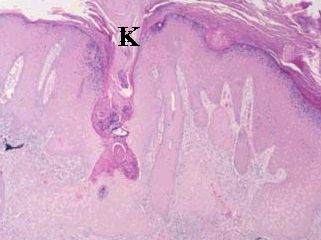
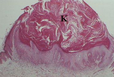
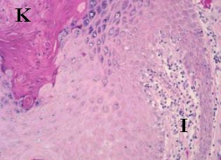
Keratoacanthoma is a rapidly growing and spontaneously resolving epidermal tumour. In the examination, you are likely to be shown either a clinical picture or a slide. 

The most common questions are:

  • What are the clinical characters of keratoacanthoma?
  • How can you differentiate keratoacanthoma from squamous cell carcinoma?


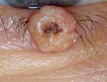
Clinically, it appears as a small erythematous nodule which grows rapidly over 3 to 4 weeks to reach a size of 2-3 cm in diameter. Central ulceration occurs commonly giving a central crater surrounded by a heaped shoulder. The lesion usually involutes over 2 to 3 months leaving an unsightly irregular scar. 


A keratoacanthoma in the lower eyelid

Histologically, the following features are seen:
  • cup-shaped lesion with a central mass of keratin 
  • acanthotic (hyperplastic) epidermis which has a sharp demarcation from the adjacent normal skin. The cells appears large and pale with small nuclei.
  • base of the lesion is well demarcated from the adjacent dermis by inflammatory response
Low magnification H&E
The specimens show presence of epidermal hyperplasia with
central crater containing keratin (K). The lesion is well demarcated 
from the surrounding (adjacent and deeper) tissues.

High magnification H&E
The slice shows keratin in the left upper corner (K). The presence of 
numerous pale large cells with small nuclei (which are the cause 
of epidermal hyperplasia) and inflammatory cells (I) in the dermis
which appear as small dark blue dots.

It is important that an adequately deep biopsy transects the lesion as the salient points in differentiating keratoacanthoma from a squamous carcinoma are the raised, rolled shoulder around the lesion and the absence of any rapidly dividing neoplastic keratinocytes blew the level fo the deepest skin appendages.

Common viva question:

  • What features are useful in distinguishing keratoacanthoma from squamous cell carcinoma?
  • How do you manage keratoacanthoma?
Return to the main page