1.
a. Right suppression
b. Left suppression
c. Exotropia (crossed diplopia)
(Worth's four dot test tests the presence of binocular vision. It consists
of four lights: two green, one red
and one white. The patient is asked to view the dots wearing red and
green filters (such that one eye sees
one red and one white light and the other sees the two green and one
white lights) and report the number
and colour of the dots he sees. Four dots indicate normal binocular
vision; two red dots indicate
suppression of the eye wearing the green filter; three green dots indicate
suppression of the eye wearing
the red filter; five dots (two red and three green) indicate diplopia
which may result from exotropia or
esotropia.)
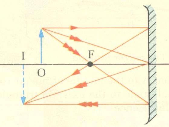
2.
a.
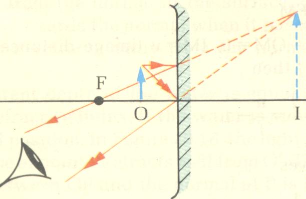
b.
F = principal point, O = object, I = image
(The centre of curvature of a concave mirror is equal to the radius of
the concave mirror whereas
the principal point of the concave mirror is equal to half the radius
of the concave mirror (r/2).
When the object is between the centre of curvature and the principal
focus, the image is real, inverted and magnified. With the object inside
the principal focus, the image moves to the other side of the mirror and
becomes virtual, erect and magnified.)
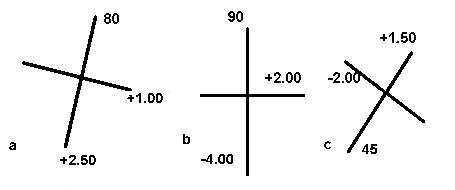
3.
a. +2.50 / -1.50 X 80 = +1.75 (spherical equivalent)
b. -4.00 / + 6.00 X 90 = -1.00 (spherical equivalent)
c. +1.50 / -3.50 X 45 = -0.25 (spherical equivalent)
(To draw the power cross remember that the power of the cylinder is
90 degrees to the axis.
The spherical equivalent is calculated by adding the value of the sphere
and half the value of the cylinder.)
4.
a. The induced prism in each eye is
5 X 1cm = 5 dioptres base up.
(Prentice's rule states that the prismatic effect is equal to the point
from the optical centre in cm multiply by the the dioptric power of the
lens)
b. 1cm X 5D X 0.2m
/ 1.0m = 1cm displacement for each eye.
(This is calculated by remembering that 1 prism dioptre deviates an
object placed 1 m away by 1 cm.)
c. Below it.
(The image is displaced downward by a a base-up prism)
5.
a. Coronal section.
b. Enlargement of the muscles in the right eyes especially
the inferior rectus.
c. Right thyroid eye disease.
d. Any of the following abnormalities may occur :
-
lid lag
-
restricted eye movement in all directions especially upgaze
6.
a. The retina and optic nerve are stimulated
with a shifting checkerboard pattern. This external visual stimulus causes
measurable electrical activity in neurons within the visual pathways. This
is called the visual evoked response (VER) and is recorded by EEG electrodes
located over the occiput. Using special computer techniques, the evoked
responses measured over multiple trials are amplified and averaged.
(With pattern-shift VER, the waveform normally appears as a straight
line with a single positive peak (100 msec after stimulus presentation).
Abnormalities in this characteristic waveform may be seen in a variety
of pathologic processes involving the optic nerve and its radiations. Pattern-shift
VER is a highly sensitive means of documenting lesions in the visual system.
It is especially useful when the disease process is subclinical for example
ophthalmologic exam is normal and patient lacks visual symptoms.)
b. Left eye which shows a significant prolonged
latency.
c. Left retrobulbar neuritis.
7.
a. The visual field shows bilateral altitudinal
field defect.
Possible causes include:
bilateral ischaemic optic neuropathy
(arteritic or non-arteritic
bilateral superior hemi-retinal artery
occlusion
bilateral superior hemi-retinal vein
occlusion
b. The visual field shows bilateral constricted
visual fields.
Possible causes include:
retinitis pigmentosa
bilateral dense laser pan-photocoagulation
advanced glaucoma
c. The visual field shows a left congruous horizontal
wedge-shaped field
defect. It is seen in lesion of
the right lateral geniculate nucleus such as
cerebrovascular accident.
8.
a. The corneal topography shows with-the-rule
astigmatism.
( The astigmatism is
shown by the typical bow-tie appearance and since the steepest part of
the
cornea is at 90 degrees
the astigmatism is with-the-rule)
b. Cutting
the cornea superiorly will flatten the cornea and therefore reduces
the astigmatism.
(provided the tension of the sutures are equally distributed and not tightened)
c.
A temporal cornea approach with small incision surgery has minimal effect
on the
corneal topography.
(Due to the small incision and the further distance between the temporal
peripheral cornea and the centre
of the cornea.)
|