Age-related macular degeneration (ARMD)
 

A dry ARMD showing drusen with
areas of RPE atrophy.

Soft drusen

Elevated macula with exudate due to 
choroidal neovascularization and 
pigment epithelium detachment. 
Age-related macular degeneration is another popular macular case. You will be expected to know the Macular 
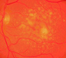
Photocoagulation Study and TAP study on photodynamic therapy (which is currently in vogue).

The macula contains drusen. In the non-exudative type there are areas of RPE atrophy. Confluent areas of atrophy 
form geographic atrophy (see picture above).

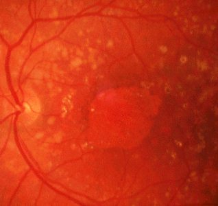
In the exudative (wet) type there may be hard exudates with or without subretinal haemorrhage.  The  macula may be 
elevated due to RPE atrophy.  In chronic exudative type, the macula contains fibrosis with RPE atrophy.

Other features to look for:

  • examine the opposite eye for similar changes. Age-related macular degeneration is always bilateral. (In younger 

  • patients, consider Doyne's disease or familial inherited drusen or Stargardt's disease).

Questions:

1. What does the Macular Photocoagulation Study show with regard to age-related macular degeneration?

2. How useful is laser treatment in treating subretinal neovascularization caused by ARMD?

3. What is the difference between classical and occult choroidal neovascularization?

4. A 63 year-old GP with a past history of wet ARMD was reviewed in your clinic. He had with him a recent paper cutting on photodynamic therapy performed by some hospital in the North. How would explain to him the theory behind this therapy?


Return to the main page


Classic choroidal neovascularization. Refer to the
fluorescein angiography below for the complete 
fluorescein angiography.

Occult choroidal neovascularization
Return to the top


A patient with a right classic subretinal neovascular membrane.