When presented with an image; the following steps are recommended:
-
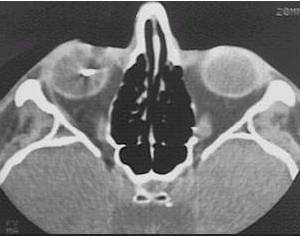
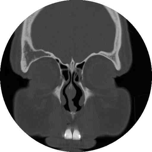
What type of image is it? CT scans or MRI scans (T1 or T2
weighted)
-
Which plane the image is in? axial, sagittal or coronal.
-
Describe the image: In ophthalmology, most of the pathology will
be focused on the globe, orbit or the brain. If it were the viva,
ask the examiner for some history.
-
Examine the following strucutres in order and always compare the
two sides for asymmetry(ies):
-
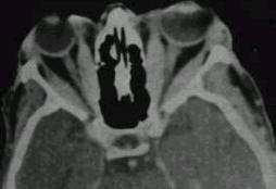
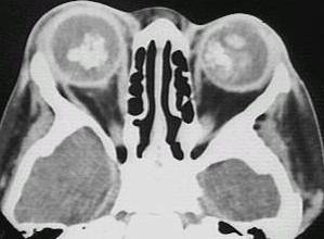
globe: eye present (enucleated eye or implant)?
lens present (aphakia)? opacities in the vitreous (blood or foreign body)?
calcification in the retina (retinoblastoma)? calcification in the optic
nerve (drusen)? any abnormal thickening of the sclera (compared with the
opposite eye, thickening suggests scleritis)?
-
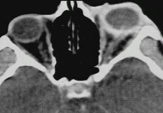
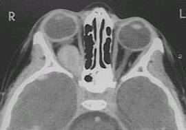
extraocular muscle enlargement (the main differetial
diagnosis include: thyroid eye disease if tendon is not involved or myositis
if the tendon is involved; other possibilities include neoplasm either
primary or secondary)?
-
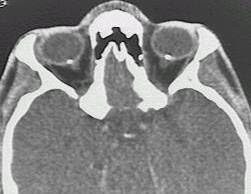
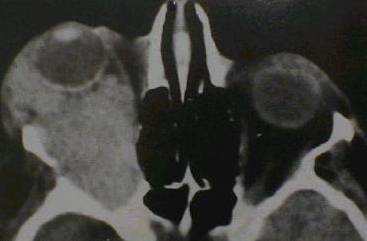
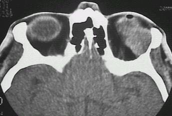
space occupying lesion in the orbit: lacrimal
fossa (lacrimal gland tumour)? intraconal lesion (optic glioma, capillary
haemangioma or optic nerve meningioma)? diffuse lesion (lymphoma, metastatic
tumour or acute infection or inflammation)? dilated superior ophthalmic
vein (carotid-cavernous fistula)?
-
bony lesion: localized bony expansion (meningioma
and osteoma in which the lesions have the same density as the surrounding
bone; fibrous dysplasia in which the bone density is reduced)? any bony
thinning next to an orbital lesion (suggesting long-standing chronic lesion)?
any lytic lesion (suggesting malignant lesions such as metastasis or myeloma)?
-
brain lesion (enlarged pituiary fossa suggests
pituitary tumour, wedge-shaped hypodense area suggests cerebral ischamia;
wedge-shaped hypodense area suggests cerebral haemorrahge)?
Click the following pictures for Questions
and Answers.
 |
 |
 |
 |
 |
 |
 |
 |
 |
 |
a |