Special thank to Dr. Squier , Consultant Neuropathologist, Radcliffe Infirmary, Oxford for providing the pictures.
Paediatric Ophthalmology: Case one

Figure 1
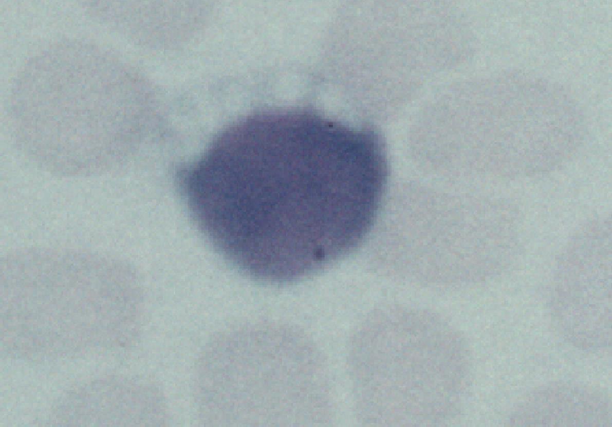
Figure 2
A 6 year old boy was referred by the paediatric neurologist for an neuro-ophthalmology assessment. He had a six-month history of deterioration of school work and appeared to have problem with his vision. Fundoscopy revealed bilateral pale discs with pigmentary changes in the periphery. Electrophysiology revealed attenuated ERG and VEP. He underwent an extensive work-up including rectal biopsy (Figure 1 is the rectal biopsy showing a neurone stained with PAS) and blood film (Figure 2).
a. What abnormalities are demonstrated in the above pictures?
The cell body of the rectal neurone (Figure 1) is stained pink due to the accumulation of the subunit c of mitochondrial ATP-synthase.b. What is the likely diagnosis?
The blood film shows vacuolated lymphocyte. (Figure 2).The diagnosis is Batten's disease of the juvenile type.c. What is the prognosis of this condition?Batten's disease (neuronal ceroid lipofuscinosis, NCL) is a group of rare autosomal recessive neurodegenerative disorders characterized by the accumulation of intralysosomal storage material. Histopathological findings include vacuolated lymphocytes, inclusion bodies in the neurones and the presence of lipofuscin and ceroid in the neurones. The precise pathogenesis of this condition is unknown but may be related to abnormalities of the mitochondrial function.
There are three main childhood types of Batten's disease with varying prognosis:Infantile
Late-infantileonset: 8 to 18 months, rapidly progressive psychomotor retardation, irritability, visual failure, hypotonia and ataxia course: myoclonic jerks, hyperkinesia, microcephaly, flat EEG by 3 years of age and death at 3 to 10 years of age Juvenile
- onset: 18 months to 4 years with epilepsy, regression, visual failure and spastic tetraplegia
- course: bulbar paresis, EEG with large polyspike response to low rates of photic stimulation, attenuated ERG and enlarged VEP, death at 4 to 10 years of age
- onset: 4 to 9 years of age, decreased vision and pigment retinopathy
- course: dementia, epilepsy, gait disorder, hallucination in older patient, attenuation of ERG and VEP, death between 15 to 30 years of age.
Click here for the questions Click here for the main page Click here for the FRCOphth/MRCOphth
/FRCS tutorials