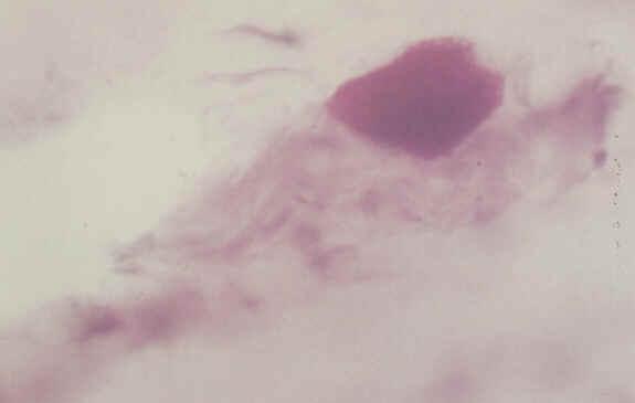
Figure 1 |
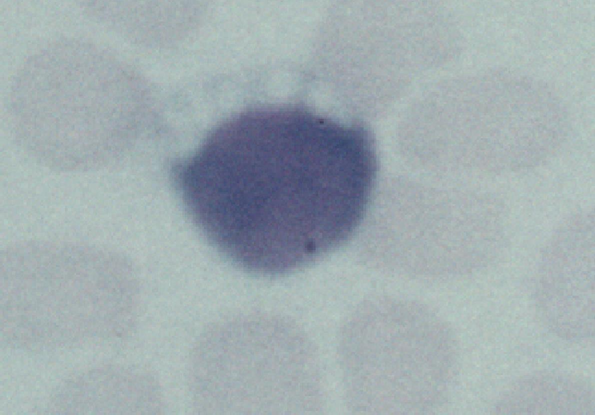
Figure 2 |
Paediatric Ophthalmology: Case one Special thank to Dr. Squier , Consultant Neuropathologist, Radcliffe Infirmary, Oxford for providing the pictures.
|
Figure 1 |
Figure 2 |
A 6 year old boy was referred by the paediatric neurologist for an neuro-ophthalmology assessment. He had a six-month history of deterioration of school work and appeared to have problem with his vision. Fundoscopy revealed bilateral pale discs with pigmentary changes in the periphery. Electrophysiology revealed attenuated ERG and VEP. He underwent an extensive work-up including rectal biopsy (Figure 1 is the rectal biopsy showing a neurone stained with PAS) and blood film (Figure 2).a. What abnormalities are demonstrated in the above pictures?
b. What is the likely diagnosis?
c. What is the prognosis of this condition?
Click here for the answers Click here for the main page Click here for the FRCOphth/MRCOphth
/FRCS tutorials