Therapeutics
Captain Cook found, in a previous unknown Australian island, a woman rubbing with a wooden stick the everted eyelids of a child. This primitive method of treating roughness of the palpebral conjunctiva seems to have a remote antiquity, and is one of the few procedures of Hippocratic ophthalmology that has persisted. Friction of the everted lid was applied by means of rough wool wrapped round a wooden spindle, the process being kept up till a thin sanguineous fluid exuded. This treatment was followed by local applications, generally containing copper.Of the more ambitious systems of treatment based on Hippocratic pathology with its crudity, coction and crisis of humours led to inactivity when it did not lead to drastic interference. In acute diseases of the eye, local remedies were avoided, and reliance placed entirely on measures influencing the humoral changes. Restriction in diet and hot foot-baths were amongst the most common, but every means that would draw the morbid humour away from the eye -- irritant gargles, cupping, venesection, cauterization of the blood-vessels in the neighbourhood of the eye, multiple incisions going down to the bone, and even trephining of the skull to evacuate the humours -- was employed. For chronic conditions, local applications containing ingredients well recognized in the more ancient civilization of Egypt were freely used -- metals and spices as well as human milk.
Alexandrian therapeutics advanced greatly on this. Local treatment for acute conditions was not only recognized but highly developed, the means employed being collyria. Unlike the modern application, the collyrium was a solid medication, made up in cakes of which gum was the basis. A fragment of a cake was dissolved in water, oil, milk of woman, urine, bile or saliva, before use. There was an endless number of these preparations, and the secret of their composition was jealously guarded. Crude lettering embossed by metal or stone stamps, of which many have been recovered in excavations, gave the name of the collyrium, fo the maker, and indications for its use. The polypharmacy of the Romans is well reflected in the composition of such collyria, the ingredients of which have been recorder. The collyrium of Hermon is reported by Celsus as containing no less than 21 substances, and the multitude of collyria recommended by him for different conditions throws an interesting sidelight on this aspect of treatment.
In addition to collyria, the Hippocratic methods of treatment were also pursued. But it was the surgery of the period that constituted a real advance. As elaborated by Galen and his commentators it supplied a rather wide range of operative treatment. Procedures for entropion and trichiasis were perfected; and an approach to the modern method of treating ectropion was made by Antyllos, as recorded by Paulus Aegineta: a triangular piece was excised from throughout the whole thickness of the lid. Operations were also evolved for lagophthlamos, tumours of the lids, "aegylops" (swelling at the inner angle), ankyloblepharon, symblepharon, pterygium and panophthalmitis. Rather complicated sutures for the cure of staphyloma of the cornea with or without resection were described by successive writers form Celsus onwards, whilst for hypopyon incision of the cornea and paracentesis were described by Galen, who also records that a certain Justus cures hypopyon by shaking the patient's head. That the Romans had a full theory and practice of cataract has already been mentioned.
After Galen superstition began to creep back into therapeutics, and with it revivals of Egyptian and Babylonian treatment by meconium faeces and similar substance. Amulets, charms and invocations figure largely in Aetius and his successors. Invoking the Deity was a usual introduction in the Arabian writings which, however, are not devoid of useful innovations, suction for cataract and peritomy in pannus being the most significant.
Astrology and its sister-study of herbs added encumbrances to the load of therapeutic measures under which ophthalmology was labouring. Towards the end of the 16th century Georg Bartisch, the father of German ophthalmology, in the first book on ophthalmology that appeared in the vernacular, devoted chapters to sorcery, white magic and black magic, though it is fair to add that Guillemeau's book -- in French -- appearing two years later (1585) is not disfigured in this manner. Easily the most crowning achievement in therapeutics during these long years of stagnation was the introduction of spectacles towards the end of the 13th century.
It was Bartisch who was responsible for the first surgical innovation that came with the Renaissance, by describing complete excision of the eye. Nearly 50 years later (1627) Fabry employed the magnet for removing a foreign body from the eye, but this procedure received no general consideration till well into the 19th century. The 17th century was indeed sterile in the field of treatment. It was left to the 18th century to introduce three epoch-making operations -- two concerned exclusively with cataract, and the third very largely with it. Early in that century Petit, basing himself on the new anatomy of cataract, described breaking up the lens in soft cataract and leaving it to absorb instead of attempting depression; and the middle of the century saw Daviel's work. But an entirely new innovation, and the opening of a chapter to which the succeeding century added greatly, was the operation for artificial pupil introduced by Cheselden (1729).
Cheselden's operation had for its object the making of an opening in the iris by a needle introduced through the sclera in cases where the pupil was blocked either congenitally, after inflammation, or after couching for cataract. To a generation which did not know of asepsis and of atropine in the treatment of the almost inevitable post-operative inflammation, the significance of the operation loomed larger than it does to us. Yet Cheselden's operation was ill-adapted to the purpose it set out to serve. Performed in eyes in which the lens was in situ, it caused traumatic cataract. Chelselden's method of introducing the needle through the sclera frequently involved injury to the ciliary body; and, most significant of all, the tear produced by a mere puncture was of transient value in most cases, any opening made contracting down or being filled with exudate before long. Attempts at improving the operation began with Sharp who, in 1740, proposed transfixing cornea and iris by one incision across the anterior chamber. Other modifications aimed at cruciform incisions and at division of the sphincter at the pupillary margin. But the operation gradually fell into disrepute and oblivion. In 1801 it was hailed as a new operation when Demours re-introduced it.
Cheselden's operation nevertheless opened a new chapter in the surgery of the iris. The modifications of his operation led to the development of iridectomy by Joseph Beer in 1798. Though a number of modifications and a variety of specially constructed instruments came on the heels of Beer's simple procedure -- carried out through a corneal incision made by the Beer knife and completed by withdrawing the iris with forceps and abscising it -- Beer's operation came to stay. Intended like Cheselden's for the formation of an artificial pupil, it led in the second half of the 19th century to the glaucoma iridectomy of von Graefe, and to its successors.
If the 18th century was successful in opening up methods for the conquest of blindness due to lens opacity and occlusion of the pupil, the 19th century groped unsuccessfully for the relief of blindness from opaque cornea. During the 18th century tentative attempts were made to resect opaque areas; Erasmus Darwin in 1795 trephined out such areas, hoping to obtain clear cornea on healing. Other attempts aimed at excising a scar and suturing clear cornea, and even at the making of windows in the sclera. But the problem which attracted most attention during the first third of the century was complete transplantation of the cornea. Successful enough on rabbits, it failed in man; the lingering discussions on the subject were revived by the suggestion (Nussabaum, 1856) that a small glass lens might be implanted in the cornea. This, too, led to disappointment; successful operation led to irritable eyes.
Closely allied to these attempts were the efforts to bring a clear part of the cornea into the line of vision. Optical iridectomy was but one of these; others aimed at iridectomy combined with the newly described operation of tenotomy, to bring the eye into a central position. Tattooing of the cornea was revived by de Wecker in 1872, after a chequered career; it had been practised by Galen, condemned by Aetius, resurrected by Guy de Chauliac in the 14th century and once again condemned by Maitre-Jan in the 18th century.
The 19th century perfected the operation of enucleation introduced by Bartisch, who incidentally had limited its indications to such massive proptosis that the eye was hideous and could not be concealed. Bonnet in 1841 and White Cooper in 1856 introduced the method of operation as it is practised today, whilst evisceration and exenteration did not come till later.
Excision as a therapeutic measure in sympathetic ophthalmia was the achievement of the second half of the nineteenth century. Sympathetic ophthalmia was first clearly indicated by Duddell in 1729, in recording that he had seen many cases in which both eyes were lost, though only one was originally injured. But it was not till nearly a hundred years later that any clear conception was developed. Demours did much in that direction, but it was Wardrop who drew attention to the fact that veterinary surgeons destroy the injured eye of a horse with lime or a nail in order that the good eye may be save. Both the writings of Demours and of Wardrop appeared in 1818, and in both the term sympathetic involvement is employed. The first comprehensive description appeared in the third edition of Mackenzie's textbook (1840), and thereafter the seriousness of the condition and its relationship to injuries and retained foreign bodies was well realized. Wardrop had advocated incision into the cornea and removal of the lens and vitreous of the injured eye as a prophylactic measure, but it was left to Prichard, of Bristol, to introduce in 1851 excision for that purpose. Only after Critchett had show, twelve years later, the ineffectiveness of excision once sympathetic inflammation had broken out, was the full value of Prichard's procedure fully appreciated. Thereafter excision rapidly replaced such methods of treatment as division of the optic nerve, of the ciliary nerves and the operation of iridectomy advocated by von Graefe.
Another procedure that was perfected during the century was the magnet operation. Dixon in 1859 deliberately incised the eye to extract a magnetic foreign body, whilst McKeown in 1874 went further; he explored the eye with the tip of a magnet introduced into the vitreous. Hirschberg a year later invented the electro-magnet.
The crowning achievement of the 19th century in ophthalmic surgery was, of course, the operative treatment of glaucoma. But it did much in plastic operations on the lids; and the introduction of asepsis and general anaesthesia was a much a boon to ophthalmic surgery as to surgery in general. The introduction of cocaine in 1884 as a local anaesthetic had of course special significance for ophthalmology.
It was also left to the 19th century to give a clear lead in the treatment of squint and of lacrimal obstruction. Both conditions had indeed been noted in antiquity, bu the conceptions concerning their nature were of the vaguest.
Squint was the evil eye of mythology and primitive folklore. in Hippocratic writings the fact that it frequently affects parents and children is clearly recognized. An early attempt at treatment is recorded in Paulus Aegineta; this consisted of wearing a mask with two perforations placed centrally before the eyes. It was argued that the squinting eye, finding vision obstructed by the mask, would assume a straight position. Fixing bright objects to the outer side of the in-turning eye was likewise attempted; it was held that the attention which these objects excited would make the eye take up a normal position. Little progress was made on this till well-nigh the 19th century. Ambroise Pare, towards the end of the 16th century, could only fall back on the method of Paulus. During the 18th century squint was regarded as the result ot malposition of the cornea or of tilting of the lens. But whilst orthodox practitioners could do nothing, the itinerant Chevalier Taylor undoubtedly put squinting eyes straight. Apparently he had discovered the fact that division of the internal rectus would sometimes straighten the eye. Surrounding his activities with much pomp and mystery, he probably performed subconjunctival tenotomies. At any rate there was always and admiring crowd to shout "a miracle." Much more significant was the work of Buffon. He recognized that the squinting eye generally had poorer vision than the fellow eye, and held that this inequality would render objects confused. His treatment was to cover the good eye, or alternatively to place a convex lens in front of it, whilst the affected eye had a plane or concave lens "in proportion to the strength or weakness of each eye."
It was well-nigh a hundred years after Taylor before surgical treatment of squint was to become common heritage. Tentatively suggested by Anthony White in 1827, and by others, the first successful operation -- a myotomy -- was performed in 1839 by Dieffenbach. Numerous modifications have followed since his day. And just as Taylor was followed by Buffon, so the surgical treatment of Dieffenbach was followed by the optical treatment of Donders, who in his classical work of 1864 showed not only the existence of hypermetropia in squint, but the frequently unequal degree of it in the two eyes and also the disturbance of balance between accommodation and convergence in hypermetropes. The fusion theory of which Javal was the main exponent, dates from about the same time.
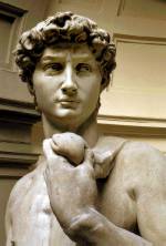
This is shown clearly in the full frontal image of David's
face, which cannot normally be
In the classic view of the statue, looking at the right
side of the face, David gazes off to
|
Lacrimal obstruction has a more prolonged and varied history. Though Galen knew the lacrimal glands, the canaliculi, and the drainage into the nose, the pathology of the lacrimal apparatus was ill understood. Under the term aegylops were included all swellings at the inner canthus; and the treatment described by Celsus, Galen and their successors was drastic in the extreme; some form of incision down to the bone and the application of the red-hot cautery was the favourite method. Among the Arabians, Avicenna may be regarded as a pioneer in treatment by probing on account of his suggestion to introduce into a lacrimal fistula probes carrying medications. The Renaissance brought accurate accounts of the lacrimal apparatus by Vesalius and Fallopius, but it was left for Stahl in the 18th century to show that the aegylops of antiquity was not an affection of the soft tissue, but the consequence of lacrimal obstruction and inflammation. Following this recognition, lacrimal affections were regarded as being either hydropsia -- when regurgitation from the sac could be obtained -- or ulcerative, when a lacrimal fistula was present. Anel in 1714 was a voice in the wilderness when he evolved a treatment for lacrimal obstruction, in which probes with an olive eminence were passed into the sac through the upper punctum whilst an astringent lotion was injected through the lower punctum by a syringe, which, like the probes, was devised by him and still bears his name.A variety of modifications were evolved. Guidethreads, for the introduction of medications into the sac, incision into the sac and catheterization through the incision; retrograde probing; and endless variety of probes; permanent implantation of tubes, were all suggested or tried at different times. Blizzard proposed the injection of metallic mercury, so that by its very weight it would clear a passage. By the beginning of the 19th century Anel's procedure had fallen into oblivion, though search still continued for the perfect method. Various attempts at cauterizing the nasal duct by silver nitrate were tried, whilst sealing the puncta was another procedure that had some vogue. It was Bowman who in 1853 re-introduced probing, employing a graduated series of instruments of comparatively large calibre. Weber advocated forcible dilatation, whilst Critchett used laminaria probes.
Though some sort of excision of the sac was practised in antiquity with its cauterization, it was not till Berlin suggested it in 1868 that excision of the sac came into ophthalmology. Two years earlier Laurence had advocated excision of the lacrimal gland, a procedure first mooted in 1843 by Bernard.
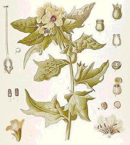
The 19th century was also responsible for the introduction of mydriatics and miotics. Mydriatics have indeed a much longer history, but their widespread clinical application only came with the second half of the century. For pain in the eyes the Greeks used opium, mandragora and hyoscyamus, a practice strongly condemned by Galen as leading to cataract and other serious complications. but Galen was not above using hyoscyamus as a cosmetic application for the blue-eyed, inducing in them a black pupil. Significant, too, is the observation by Pliny that anagallis is used for dilating the pupil before couching operations; this procedure is not mentioned anywhere else in the old literature, and the reference is all the more puzzling as anagallis has no mydriatic effect; but in accuracy of details the garrulous Pliny is never too reliable. Whatever vogue mydriatics may have had in Greece and Rome they lost during the succeeding centuries. The rediscovery came towards the end of the 18th century. Though John Ray, the Father of Natural History in England, recorded in 1686 his observation that a belladonna leaf applied to a small abscess near the eye had caused dilatation of the pupil, it was not till another century had passed that mydriatics received any attention. This came with the reports of three different observers (Daries, Loder, Reimarus), who independently recorded the mydriatic action of belladonna. Loder in 1796 and Reimarus in 1797 advocated its use to facilitate cataract extraction, a practice that was adopted in England by Paget of Leicester in 1801 and John Cunningham Saunders in 1809. Himly in particular did much to study systemically the use and possibilities of mydriatics in ophthalmology, yet it was not till 1831 that atropine was isolated.
|
Hyoscyamus from which hyoscine is obtained. |
The first half of the 19th century was satisfied with general treatment of iritis; the sue of mydriatics for this condition, though indicated as early as 1805 by Schmidt, did not gain any widespread acceptance; von Ammon in 1835 could still advise against their use in acute cases; and whilst Desmarres in 1847 strongly recommended belladonna in his textbook, his German translator could only report that the drug causes an increase in the inflammation. It was largely through the advocacy of von Graefe in 1856 that atropine came to occupy its place in modern ophthalmology. No doubt the dispute over the applicability of atropine was in part the consequence of the non-recognition of acute and subacute glaucoma as distinct from iritis. Even when that recognition came, the deleterious action of atropine in glaucoma was not appreciated; that only came with von Graefe in 1868.Miotics have a much briefer history. The pharmacology of the calabar bean was studies at Edinburgh in 1846 and 1855. Fraser, subsequently Professor of Pharmacology at that University, showed its miotic effects in 1862. A year later Argyll Robertson demonstrated its effects on accommodation. In the same year von Graefe studied its antagonistic action to atropine and employed it to facilitate iridectomy in non-inflammatory glaucoma. It was during the succeeding decade, after von Graefe had shown the danger of atropine in glaucoma, that miotics began to be employed as therapeutic agents. Eserine was isolated in 1864 and pilocarpine in 1875.