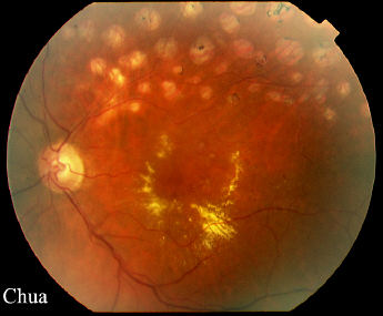
| History: This 58 year-old man suffered from a
superotemporal retinal branch vein occlusion 12 months ago. The
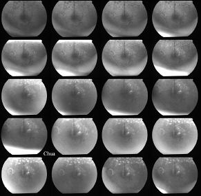
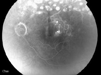
vision at presentation was 6/9. Five months after the initial presentation he developed a vitreous haemorrhage attributed to peripheral neovascularization, sectorial pan-photocoagulation was performed (732 spots X 200um X 0.1 sec X 200-250 mW) with resolution of the new vessels. At the 9-month follow-up, his vision decreased from 6/9 to 6/24 due to macular oedema with circinate exudate. Fluorescein angiography showed vascular leakage from microaneurysms. Focal argon laser was performed (48 spots X 100um X 0.1sec X 90-110mW) aiming at the microaneurysms and area of oedema but avoiding the fovea. At 3 month follow-up, the oedema resolved and the vision improved to 6/12 (click on the picture for a magnified view). |
|||
 a a |
|||
|