.
The most common scenario in the examination is young
female with history of multiple sclerosis. However, it can also be
seen in older patients with cerebrovascular accident.
The main feature of this condition is impaired adduction. A favourite
question is the site and side of the lesion (see question
below).
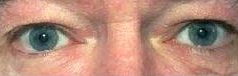
In unilateral case, the affected eye shows failure (or
impaired) adduction (failure of conjugate eye movement). The abducting
eye shows jerk nystagmus with the quick phase towards
the opposite side (this is called ataxic nystagmus but may not be
obvious and can be absent). The horizontal saccade
is abnormal with the affected eye lagging behind the normal eye. The
vertical saccade and convergence are normal.
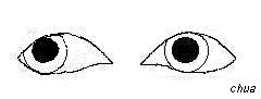
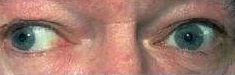
Left saccade abnormality.
This may be the only sign present in patient
with recovered internuclearophthalmoplegia.
|
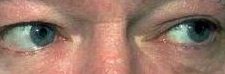
In the examination:
-
there may be strabismus in the primary position
-
the internuclear ophthalmoplegia may be bilateral with or
without asymmetry
-
if ask for further examination, mention that you would like
to examine the patient for signs of demyelination:
-
fundal examination for pale disc (from previous optic neuritis
which may be in either eye)
-
afferent pupillary defect (again from previous optic neuritis)
-
cerebellar signs (for example scanning speech, disdianochokinesia,
intentional tremor,
past-pointing in finger nose test)
|