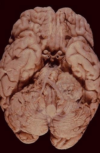
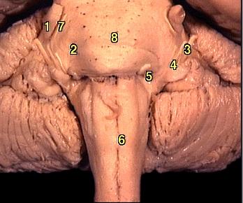
Cerebellopontine angle lesion
 

Post-mortem specimen of showing a left acoustic 
neuroma.

Anatomy of the cerebellopontine angle showing the 
relationship of the various cranial nerves.
1.Sensory root of trigeminal nerve 
2.Pons 
3.Vestibulocochlear nerve 
4.Facial nerve 
5.Abducent nerve 
6.Medulla oblongata 
7.Motor root of trigeminal nerve 
8.Basilar sulcus 

There are several conditions which can give rise to cerebellopontinge lesion (for example, acoustic neuroma, 
meningioma and metastasis) but in the examination the most common cause is acoustic neuroma.

On the affected side, there is jerk nystagmus with the fast phase to the affected side (if the nystagmus is cerebellar; 
but in the early stage the initial nystagmus may be of vestibular origin with the fast phase to the unaffected side). 
The ipsilateral fifth nerve function is impaired (motor, sensory and reflexes ie. corneal and jaw are involved but in 
the early stages the corneal reflex alone may be involved). The sixth and seventh nerves are involved (but this may 
be mild in the early stages). The hearing on the ipsilateral side is impaired.

Further examination:

  • mention you would like to examine for other cerebellar signs
  • take a family history as patients with type II neurofibromatosis are at risk of developing bilateral acoustic 

  • neuroma.

Questions:

1. What is the different between acoustic neuroma and vestibular schwannoma?

2. What does the MRI scan below show?
 

3. The following are the two histological appearances of schwannoma. Which one is Antoni A pattern and which one is Antoni B pattern?
 

Figure 1

Figure 2

Return to the main page