Common visual field defects 

In the final MRCOphth, most cases of visual field defects are related to neurological disorders as shown below. 
Occasionally you may be asked to demonstrate visual field defects in patients with retinal problems and the most 
common field defects and their underlying conditions are:
  • tunnel vision in advanced retinitis pigmentosa and advanced glaucoma
  • altitudinal field defect in ischaemic optic neuropathy and branch retinal artery occlusion
  • arcuate scotoma in glaucoma
The image below is the MRI scan of a brain showing the optic nerves and the visual pathway. Please click on 
the number for the visual field defects and questions:

 
Layer 2Layer 3Layer 4Layer 5Layer 6Layer 7 
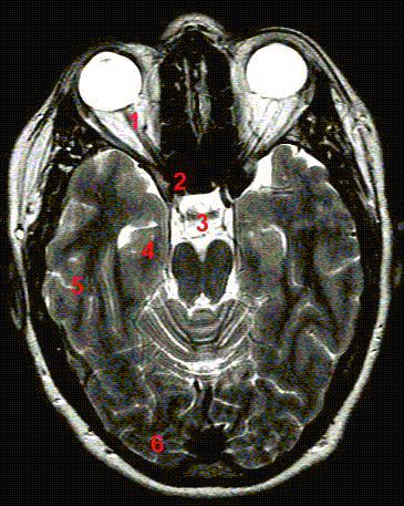
1. Central scotoma resulting from inflammation of the 
    optic disc

2. Junctional scotoma

3. Bitemporal hemianopia resulting from a lesion around 
    the optic chiasm

4. Incongruous homonymous hemianopia resulting from 
    a lesion in the optic tract

5. Homonymous quadrinopia resulting from a lesion in
    the temporal lobe

6. Homonymous hemianopia resulting from a lesion in 
    the occipital lobe.

Return to the main page