Figure 1 |
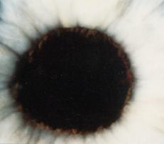
Figure 2 |
Eyelids and Anterior Segment: Case two
|
Figure 1 |
Figure 2 |
This 65 year old woman complained of a right blurred vision. There was no history of trauma and she was otherwise in good health. On examination, her right visual acuity was 6/60 and that of the left is 6/6. The appearance of her right eye on slit-lamp is shown in Figure 1. Figure 2 was taken two weeks later.a. What is the cause of her blurred vision?
Spontaneous hyphaema caused by vascular tufts (microhaemangiomas) of the iris.Microhaemangiomas tend to occur in late adulthood, appearing after the fifth decade of life. They contain clusters of coiled, thin-walled vessels and have a mulberry appearance at the pupillary margin. Due to their small size and tendency to be obscured by pigment, they are best visualised with high magnification slit lamp microscopy.
Fluorescein angiography may be needed to demonstrate them conclusively. Spontaneous hyphaema due to vascular tufts tend to absorb rapidly, with only transient visual disturbance or raised intraocular pressure. There is no association with any systemic illnesses.
b. What could be the other causes as seen on Figure 1?
Causes of spontaneous hyphaema include:
- vascular abnormalities
rubeosis iridis
vascular tufts
vasularised pupillary membrane- inflammatory processes
herpes zoster
Fuch's heterochromic iridocylitis- vascular erosion
juvenile xanthogranuloma
iris naevus or melanoma- haematologic disorders
anticoagulants
thrombocytopaenia- late surgical complications
cataract or glaucoma surgery
laser iridotomy
| Click here for questions | Click here to return to the main page | Click here for MRCOphth/FRCS tutorials |