Medical Retina: Case three
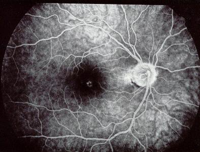
Figure 1 |
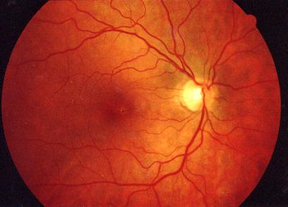
Figure 2 |
This is the fluorescein angiography of a 56 year old woman who developed a distorted right vision a few hours after observing a solar eclipse. She complained of distorted right vision and the visual acuity was 6/12 in this eye.a. What does the fluorescein angiography show?
The fluorescein angiography shows a small area of hyperfluorescence in the fovea. This transmission defect is caused by damage to the retinal pigment epithelium due to solar retinopathy
b. What is the mechanism of this condition?
Solar retinopathy occurs as a result of retinal injury from sun-gazing. Two mechanisms are thought to be responsible:
photochemical damage (from short visible wavelengths)
thermal damage (damage is related to the duration of glazing and the size of the pupil)
Histology of solar retinopathy revealed injury to the retinal pigment epithelium with detachment and necrosis. The damage to the photoreceptors is usually minimal.c. What is the natural history of this condition?
In solar retinopathy, the vision is affected 1 to 4 hours after sun-glazing. The typical complaints being decreased or distorted vision, photophobia and eyeache.
In acute stage, there is a yellow foveal lesion with surrounding macular oedema. Over time, the swelling resolves leaving behind hyperpigmentation in the macula (Figure 2). The visual acuity usually improves to 6/6 to 6/12 although distortion and a small area of scotoma may be present.
Click here for the answers Click here for the main page Click here for FRCOphth/MRCOphth
/FRCS tutorials