|
|
|
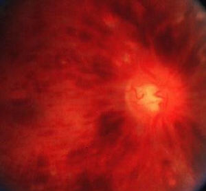
There are retinal haemorrhages (dot and blot haemorrhages and flame haemorrhages) in all quadrants. The veins are tortuous and engorged. There may be multiple cotton-wool spots. The optic disc may be swollen in the acute stage. In chronic cases, the signs are less florid and do not omit to look for disc collaterals, macular oedema and peripheral neovascularization. (Note: in chronic cases, the signs may be similar to diabetic retinopathy.) Also look for:
and rubeosis iridis and hypertensive changes in the contralateral eye) |
Questions:
1. What are the risk factors for central retinal vein occlusion?Answer
2. What are the risk factors for neovascularization in central retinal vein occlusion?
