|
||
|
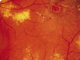
In the examination, most cases of Coat's disease seen are of the less severe types in which there are peripheral features of Coat's disease. The severe types of Coat's disease in which there are leucocoria, strabismus and extensive exudative retinal detachment may appear in other sections such as ocular motility examination. Most patients are of the young age and usually male but occasionally you may get older patients with adult-onset Coat's disease. The macula shows hard exudate with oedema. The peripheral
retina has telangiectatic and tortuous blood vessels (which
Look for:
|

Questions:
1. When can you see light bulb changes in Coat's disease?