Retinitis Pigmentosa
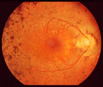
Retinitis pigmentosa showing the typical bone spicules pigmentary changes. |
| There are multiple bone-spicules
hyperpigmentation in the peripheral retina. In advanced cases,
the posterior pole is also involved. The retinal vessels are attenuated and the optic disc is pale. The macula may show cellophane, atrophic or cystoid maculopathy. The condition is bilateral. Other features to look for:
may also have deafness and pigmentary changes in the retina) |
Questions:
1. Which conditions may simulate retinitis pigmentosa?