Retinoschisis
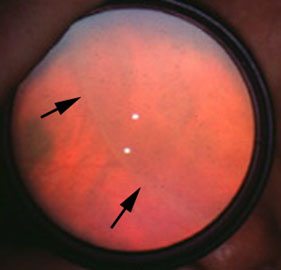 |
This is a common case for indirect ophthalmoscopy
using a 20D.
There is a dome-shaped, smoothly elevated transparent lesion in the peripheral retina. Most commonly seen in the upper temporal quadrant. There are holes on the lesions. Other signs:
|
Questions:
1. How does retinoschisis develop?