Retinal Detachment
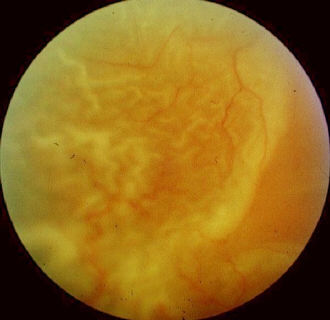 |
| Note: The majority of the cases has treated retinal
detachment possibly with proliferative vitreoretinopathy
(PVR). Otherwise, the patient may be an in-patient awaiting surgery
(the wearing of hospital pyjama may provide the clue). Another favorite
case is long-standing retinal detachment usually with
watermark.
In treated retinal detachment, there is/are area(s) of chorioretinal atrophy with indentation from cryotherapy and explant. Examine the conjunctiva and comment if the explant is secured. Look for wrinkling of the retinal surface, vessel tortusosity and retinal folds suggesting the development of PVR. Examine the opposite eye for lattice degeneration and cryo/photocoagulation scars. Note presence of other predisposing factors:
|
Questions:
1. List the risk factors for retinal detachment