Choroidal melanoma
 |
| Choroidal naevus or melanoma may be easily missed
if you were asked to examine with an direct ophthalmoscope especially if
the lesion is only slightly pigmented. Remember to look for this if the
optic disc, macula and retinal vessels are normal.
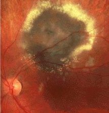
The lesion appears as an elevated, pigmented mass. There may be orange pigment (lipofuscin) on the tumour. Look for any retinal detachment around it. In melanoma treated with plaque, the surrounding area shows atrophy or surrounded by hard exudate (see the picture on the left). Other signs:
|
Questions:
1. What treatment options are available for choroidal
melanoma?