Blunt trauma
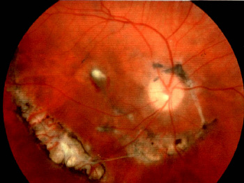 |
| The posterior pole contains white lines (exposed sclera)
which are concentric to the optic nerve. The edges of the lines may be
hyperpigmented due to RPE hyperplasia. There may be retinal oedema or haemorrhage due to subretinal neovascular membrane. There may be other associated signs such as macular hole.
The peripheral retina may have chorioretinal atrophy due to retinal
Other sings
|
Questions:
1. How does choroidal rupture occur?