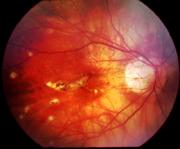
History: A 43 year-old myopic woman was referred
by her optician because of reduced right vision
and multiple atrophic scars in the macula. Her
previous retina examination 2 years ago was
normal. The vision was 6/24 in the right eye
and 6/9 in the left eye. There was no vitritis. The
appearance is suggestive of the condition punctate
inner choroidopathy (PIC).
This condition is characterized by:
- moderate myopia
- female sex
- multiple, yellow-white lesions of the inner
choroid and retina confined to the posterior
pole, and that after resolution
leave atrophic pigmented scars
- frequent serous detachment of the retina that resolves
spontaneously
- absence of vitritis or anterior uveitis
- bilateral ocular involvement common
- negative histoplasmin skin test (70%)
- choroidal neovascularization in 40% of eyes
- relatively good visual prognosis with 50% retaining
normal acuity
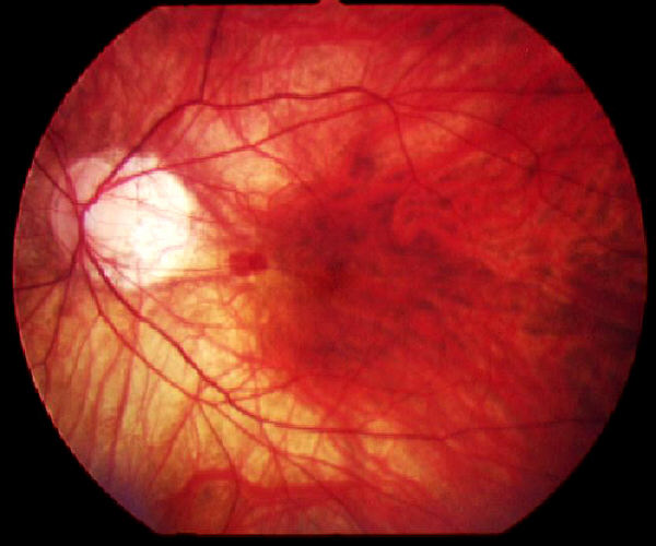
The vision remained unchanged in her right eye in
subsequent follow-up and no signs of subretinal
neovascular membrane. In one of the visits she was
noted to have a blot haemorrhage in
her left eye but her blood pressure and blood tests
including glucose were normal and the
intraocular pressures in both eyes were normal. The
haemorrhage resolved spontaneously.
|