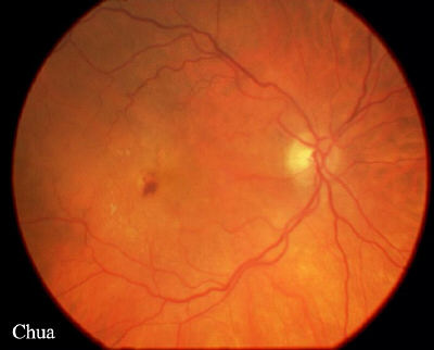
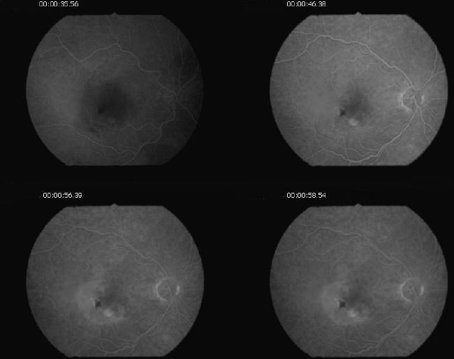
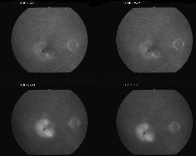
| History: A 85 year-old woman with a known history of dry age-related macular degeneration in both eyes was referred by her optician because of a 2-month history of deteriorating right vision and a small area of haemorrhage. On examination her vision was 6/36 in the right eye (6/9 two years ago) and 6/12 in the left eye. Fundoscopy showed the presence of a blot haemorrhage and slightly raised macula in the right eye. The left eye showed only multiple drusens. The FFA showed classic subretinal neovascular membrane and pigment epithelium detachment (PED). Photodynamic therapy was not performed because of the presence of the PED. | |||
 |
|||
 |
|||
 |
|||
|