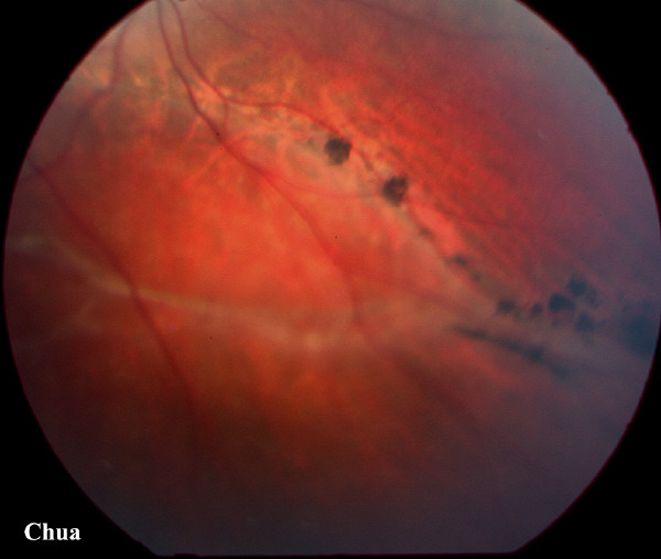
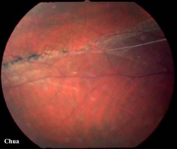
| History: A 50 year-old man was referred by his optician because of pigmented areas in the right inferior retina. The patient was asympatomatic and the vision was 6/6 in both eyes. Fundoscopy revealed pigmented 'watermark' and a right inferior retinal detachment. As the macula was not involved, no surgery was undertaken. The patient was reviewed at nine-monthly interval but was asked to contact the eye unit should he experience any right visual distortion. The pigmented areas are caused by the proliferation of retinal pigmented epithelium that have escaped through the retinal tear. Because of gravity, chronic asympatomatic retinal detachment is uncommon in superior retinal detachment. | |||
 |
|||
 |
|||
|