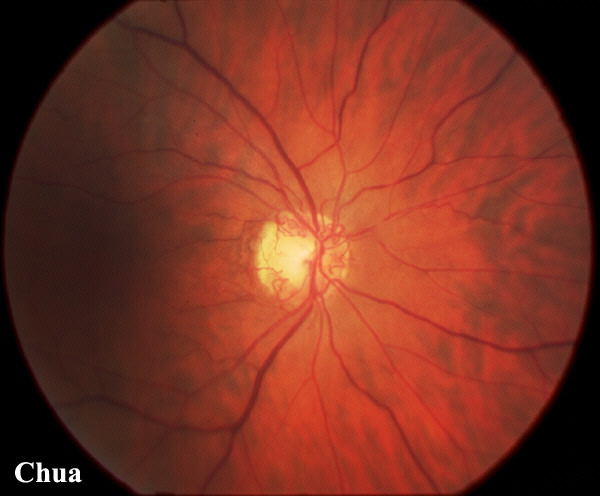
| History: A 60 year old hypertensive man developed a right cilioretinal artery occlusion which reduced his vision to 6/36. When seen two months later, the right optic disc showed multiple collaterals and the vision was 6/18. The collaterals often appear as tortuous vascular loops and are due to opening of the vascular channels between the retinal and the ciliary vessels. They are also called optociliary vessels. They may sometimes be mistaken for new vessels but unlike new vessels, they are of larger calibre and do not bleed. Fluorescein angiogram in collaterals show no leakage as in new vessels. | |||
 |
|||
|