| History: This 73 year-old woman presented to the eye casualty with a sudden onset left central scotoma of 2 day duration. The visual acuity of the affected eye was 6/18. Her past medical history consisted of angina under medical control. Fundal examination revealed an area of paleness and retinal thickening corresponding to the distribution of a branch of the lower branch retinal artery. The appearance was consistent with a branch retinal artery occlusion. No emboli was seen in either fundi. A full blood count and ESR were normal. There was no carotid bruit, her pulse was regular and cardiovascular examination revealed normal heart sounds. The carotid ultrasound did not show any plaque and the echocardiogram was negative for thrombus. No further treatment was carried out as the patient was already on 75 mg aspirin for her angina. The oedema resolved with time and the scotoma improved with time. The visual acuity improved to 6/9 | |||
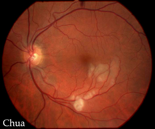
An area of paleness and retinal oedema corresponding to the distribution of a branch of the inferior retinal artery. |
|||
|