| History: A 75 year old hypertensive
man suffered from a right branch retinal vein 6 months ago. In the follow-up
clinic, his vision remained poor at 6/36. Fundoscopy revealed macular oedema.
The area of cystoid macular oedema is clearly shown by the OCT (optical coherence tomography) scan. A fluorescein angiography was carried out which showed areas of ischamia in the foveal region without leakage. Argon laser was not performed. OCT is useful in showing retinal thickening and oedema especially in patients where medial opacities make macular view difficult. However, it does not give information about the circulation. In deciding if macular oedema secondary to branch retinal vein occlusion is suitable for laser treatment, fluorescein angiography is essential to exclude ischaemia and to localize area(s) of leakage. |
|||
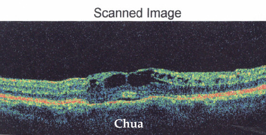
The OCT scan shows retinal thickening and cystoid spaces. |
|||
|