Relative afferent pupillary defect
 
.

Equal pupil sizes 

Normal direct and consensual light reflexes.

Left relative afferent pupillary defect.

Normal direct and consensual reflexes.
Note: The arrows represent the light.
.
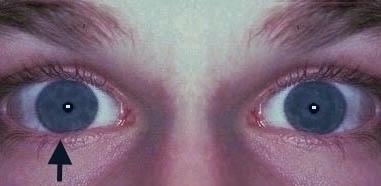
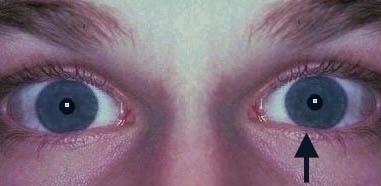
This is a common case in pupillary examination. Always suspect this if there is no anisocoria.

The direct and consensual pupillary responses to light are normal. The swinging light test shows abnormal light response of the affected eye (initial dilatation followed by constriction). For example, if the left eye were abnormal, both pupils constrict when the light is shown into the right eye. When the light is swung to the left eye, both pupils dilate. When the light is swung back to the right eye both pupils again constrict. This reaction indicates a defect in the afferent pupillary fibres from the left eye. The near reflex is normal.

Further examination:

  • tell the examiner that you would like to examine the fundus of the affected eye. The most common physical signs would be optic atrophy. Other possibilities include advanced glaucoma, retinitis pigmentosa, old central retinal artery or vein occlusion.

A patient with a left relative afferent pupillary defect.

Questions:

1. What is the significance of a relative afferent pupillary defect?

2. Would you expect a patient with bilateral optic neuritis to exhibit relative afferent pupillary defect?

3. How can you assess the amount of afferent pupillary defect?

4. Is it possible to detect relative afferent pupillary defect if one of the eye were dilated (also see diagrams below)?

Return to the main page


Fig.1 A patient with a right dilated and unreactive pupil.
The swinging flash test shows abnormality of the left eye.
(Note the dilatation of the left eye when the light is swung to the left.)


Fig. 2 A patient with a right dilated and uncreactive pupil.
The swinging flash test shows abnormality of the right eye 
(Note dilatation of the left eye when the light is swung to the right).