Figure 1 |
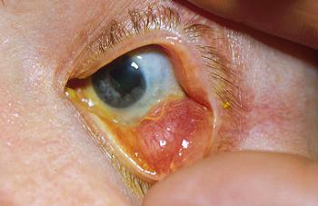
Figure 2 |
Eyelids and anterior segment: Case seven
|
Figure 1 |
Figure 2 |
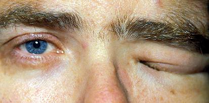
This 50 year-old man has a long history of difficulty in opening his left eye (Figure 1). Figure 2 shows the appearance of the left eye when it is manually opened.a. What physical signs are present and what is the most likely diagnosis?
Figure 1 shows swelling of the left eyelids with ptosis. Figure 2 shows lobulated lesions in the left lower lids and ectropion uvea.Left plexiform neurofibroma caused by neurofibromatosis type I.
It is composed of a proliferation of Schwann cells and fibroblasts embedded in endoneurial collagen. On palpation, the lesion is classically described as having the feel of a bag of worms.
b. What other ocular signs may be seen in this patient?The following signs are seen in neurofibromatosis type I:
- Enlarged corneal nerves
- Lisch nodules
- Glaucoma
- Astrocytic hamartoma
- Afferent pupillary defect or optic atrophy from optic nerve glioma or meningioma
- Proptosis from orbital neurofibroma or optic nerve glioma or meningioma
- Pulsating proptosis from absent sphenoid bone
c. How successful is surgical correction of this condition?Mechanical debulking is performed in mechanical ptosis resulting in loss of vision. However, due to the highly infiltrative nature of the tumour complete excision is difficult and the tumour recurs easily.
| Click here for the questions | Click here for the main page | Click here for FRCOphth/MRCOphth
/FRCS tutorials |