Figure 1 |
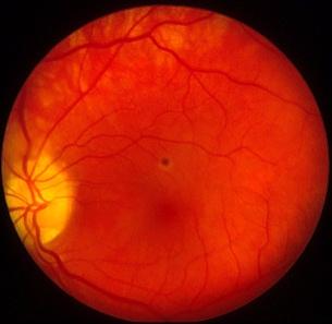
Figure 2 |
| Medical Retina & Posterior Segment: |
| Case 25 |
|
Figure 1 |
Figure 2 |
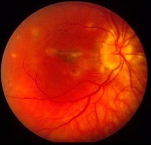
| This 29 year-old woman presented to the eye
casualty with a one-week history of distorted vision in both eyes. Her
visual acuity was 6/12 in the right eye and 6/9 in the left with a myopic
correction of -2.75 and -3.25 respectively. Slit-lamp examination of the
anterior segment was normal and there was no vitritis. However, fundoscopy
revealed multiple small white lesions. The blood tests including ACE, VDRL/TPHA
and autoantibodies were normal. Her past medical history was unremarkable.
The above pictures were taken five weeks later.
a. What is the most likely diagnosis? b. What is the differential diagnosis? c. What complication may occur with this condition? d. How would you treat this condition? |
| Click here for the answers | Click here for the main page | Click here for MRCOPhth / FRCS tutorials |