Figure 1 |
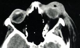
Figure 2 |
Eyelids and anterior segment: Case six
|
Figure 1 |
Figure 2 |
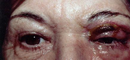
This 46 year old presented with a two-day history of painful right eye and this appearance. A CT scan was requested as shown.a. What is the diagnosis?
Necrotizing fasciitis. There is necrosis of the upper lid with periorbital swelling. The CT scan shows involvement of the deep subcutaneous tissue.This is a potentially fatal infection of the deep subcutaneous tissue. In the eyelid, it typically presents as pre-septal cellulitis and rapidly progresses to necrotizing cellultic lesion. The necrosis is caused by septic thrombophlebitis of dermal vessels caused by the bacteria.
The patient is usually toxic and may die of septicaemia if not treated early.
b. What is the most common pathogens involved?The two most common pathogens are:
- Beta-haemolytic streptococci of Lancefield's group A (Strep. pyogenes) or group C and G
- Staph. aureus
c. How would you manage this condition?This is a medical emergency and the patient must be admitted for treatment.The antibiotics of choice are high doses of benzyl penicillin (3MU 4 hourly) to achieve adequate penetration of the necrotic tissue and flucloxacillin to cover the staphylococcus.
Debridement of the necrotic tissue. Some advocate early debridement but other perform this only if the edge of the necrotic tissue continue to advance after 24 hours of initiating antibiotics (indicating failure of antibiotic to penetrate the necrotic tissue).
| Click here for the questions | Click here for the main page | Click here for FRCOphth/MRCOphth
/FRCS tutorials |