Figure 1 |
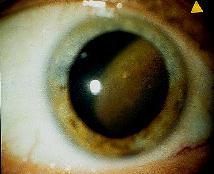
Figure 2 |
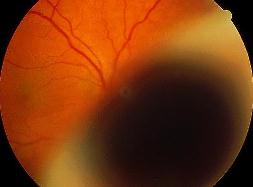
Figure 3 |
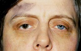
This 45 year old woman was referred to the clinic because of recent visual disturbance in the right eye. Her visual acuity was hand movement in the right eye and 6/6 in the left eye. Figure 1 shows her facial appearance (the right pupil had been dilated) and figure 2 the slit-lamp appearance of her right eye.a. What do the pictures show?
Figure 1 shows pigmentation over the right temple and heterochromic iridis (the right iris being darker than the left)Figure 2 shows a pigmented lesion that extends into the vitreal cavity
b. What is the most likely diagnosis?Oculodermal melanocytosis (naevus of Ota) with associated uveal melanoma.Naevus of Ota is a form of congenital melanosis characterized by hyperpigmentation of the affected tissue as a result of an increased numbers of heavily pigmented melanocytes within the tissue. The affected areas include the facial skin, eyelids, conjunctiva, sclera, episclera and the uveal tract.
The condition is more common in Asians than black or white. Uveal melanoma is an occasional complication of this condition.
c. How would you manage this patient?Detailed fundal examination and the use of ultrasound are important to establish the nature of the lesion. (Figure 3 shows the lesion in figure 2 arises from the ciliary body and extends posteriorly, ultrasound of the lesion showed features of melanoma with acoustic hallowness and choroidal excavation)General physical examination is important to exclude the possibility of a primary carcinoma (breast in females) as well as metastasis (uveal carcinoma has a predilection for liver). Liver function tests and liver ultrasound are useful to detect hepatic metastasis.
Treatment option will depend on the size of the lesion. In this patient, the tumour was enulceated as it was too large to be treated with radiotherapy or other mode of treatment.
| Click here for the questions | Click here for the main page | Click here for FRCOphth/MRCOphth
/FRCS tutorials |