Figure 1 |
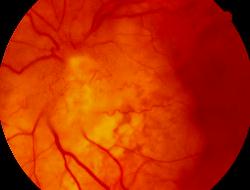
Figure 2 |
Medical Retina: Case fourteen
|
Figure 1 |
Figure 2 |
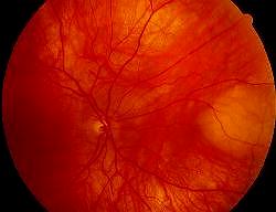
This 56 year-old woman complained of decreased left vision. She was on chemotherapy for breast carcinoma. Her right and left fundi are shown in figure 1 and 2 respectively.a. What do the pictures show?
Figure 1 shows a yellowish lesion in the nasal aspect of the right retina. Figure 2 shows left disc swelling and yellowish lesions arising from the optic disc.b. What is the cause of her decreased vision?Metastatic cancer involving the left optic disc.Metastatic tumour in the choroid (figure 1) is the most common ocular tumour in adult but rarely causes visual problem.
However, optic disc involvement (figure 2) is uncommon. It is usually caused by direct extension of a juxtapapillary choroidal metastasis but occasionally the optic disc alone is affected alone.
The associated findings include optic disc oedema, dilated and tortuous retinal vessels, afferent pupillary defect and less commonly retinal artery or vein occlusion. The visual prognosis is poor.
c. Apart from metastases, in what way can the vision be affected by malignancy?Vision may be affected by cancer associated retinopathy (CAR) syndrome. It is a type of paraneoplastic retinopathy characterized by photoreceptor degeneration with insidious loss of vision over several months. Initially, the retina may appear normal but later on there is thinning of the retina and vessels attenuation. It is classically associated with oat cell carcinoma of the lung.
| Click here for the questions | Click here for the main page | Click here for FRCOphth/MRCOPhth
/FRCS tutorials |