 |
Medical Retina & the Posterior Segment:
Case nineteen
|
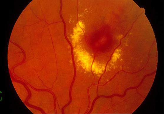
This 65 year-old woman was seen in the eye casualty because of a distorted vision in her left eye. Her visual acuity was 6/6 in both eyes. She had no diabetes mellitus but was on medication for hypertension.a. What is the diagnosis?
Macroaneurysm.The picture shows an area of haemorrhage in and around a branch of the superior retinal artery surrounded by a circinate lipid exudation.
Retinal macroaneurysm is caused by abnormal dilatation of the retinal arteriole within the first three orders of bifurcation. Hypertension is seen in 75% of patients and the ratio of women to men being affected is 3:1.
b. What complications can occur with this condition?Macroaneurysm causes visual problems through:
- Haemorrhage. This may be intraretinal, subretinal or vitreous haemorrhage
- Leakage of plasma. This can lead to cystoid macular oedema and serous macular detachment.
- Occlusion of the distal retinal artery. The site of macroaneurysm can give rise to thrombus leading to emboli in the distal retinal artery
c. How do you manage this condition?Treatment is not needed if the macula is not involved as the macroaneurysm can thrombose leading to spontaneous closure and resolution of the haemorrhage and oedema.However, if the macular vision is involved focal argon laser to the periphery of the macroaneurysm can reduce the oedema. Direct laser application is not advised as it can lead to distal retinal arterial occlusion.
| Click here for the questions | Click here for the main page | Click here for FRCOphth/MRCOphth
/FRCS tutorials |