 |
Eyelids and the Anterior Segment:
Case thirty five
|
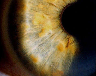
This 25 year-old man was seen in the ophthalmic genetic clinic and was found to have this iris appearance.a. What is the diagnosis?
Lisch's nodules.There are multiple, well-circumscribed and raised yellowish-brown nodules on the iris. This patient was seen in the clinic because his father is known to suffer from neurofibromatosis type 1.
b. What is the chance of his offspring having this condition?50%. Lisch nodules occur in patient with neurofibromatosis type 1. This condition is autosomal dominant and the abnormal gene has been traced to the long arm of chromosome 17.c. What is the histopathology of this condition?
Lisch's nodules are iris hamartoma containing melanocytes. The nodules are rarely seen in infants but become more prominent as the patients grow. They are seen in 90% of adult patients with type 1 neurofibromatosis.
d. What other conditions may be present in this patient?Other ophthalmic findings:Facial appearance
- eyelids: plexiform neuromas causing mechanical ptosis
- cornea: prominent corneal nerves
- glaucoma
- retina: astrocytic hamartoma
- optic disc: glioma causing optic atrophy with or without proptosis
- orbital abnormality: absent sphenoid causing spheno-orbital encephalocele and giving rise to pulsatile proptosis
Systemic conditions:
- facial asymmetry such as hemi-hypertrophy
- skin: cafe au lait spots, axillary freckles and neuroma
- intracranial tumours such as meningioma, ependymoma and glioma
- other tumours: phaeochromocytoma, medullary carcinoma, cardiac rhabdomyoma.
- congenital bony defects: scoliosis
| Click here for the questions | Click here for the main page | Click here for FRCOphth/MRCOphth
/FRCS tutorials |