Figure 1 |
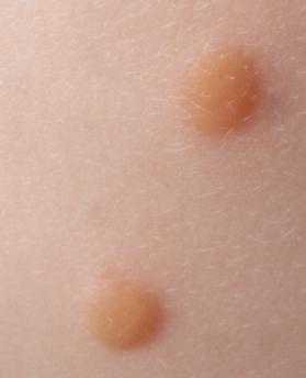
Figure 2 |
Paediatric Ophthalmology: Case eleven
|
Figure 1 |
Figure 2 |
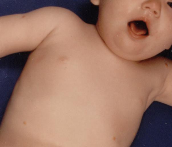
This eleven month old baby girl was referred by the paediatrician for an ophthalmic examination because of this skin appearance (figure 1 and figure 2). Histology of the lesion showed giant cells.a. What is the diagnosis?
Juvenile xanthogranuloma.The condition consists of benign proliferation of histiocytes of unknown origin. It is a disease of childhood (typically affect children less than one year old). The skin lesions appear as multiple small yellowish nodules and tend to occur in the head, neck and extremities. The histology is characteristic with non-caseating histiocytic infiltration and Touton giant cell (multinucleated cells surrounded by cytoplasm laden with lipid). The condition usually regresses spontaneously.
b. What ophthalmic sign(s) would you be looking for?Juvenile xanthogranuloma can affect the conjunctiva, iris and the ciliary body. Most ophthalmic signs relate to iris involvement and can give rise to:
- iris tumour which has a tendency to cause spontaneous hyphaema
- glaucoma
- uveitis
- heterochormic iridis
c. How do you manage this condition?The skin condition regresses spontaneously and does not require treatment.
Ocular involvement is usually treated conservatively as the lesion tend to regress. If there were recurrent hyphaema or uveitis, a short course of topical and systemic steroid can be used to induce remission. If this fail, radiotherapy can be useful.
| Click here for the questions | Click here for the main page | Click here for FRCOphth/MRCOphth
/FRCS tutorials |