 |
Eyelids & the Anterior Segment:
Case twenty three
|
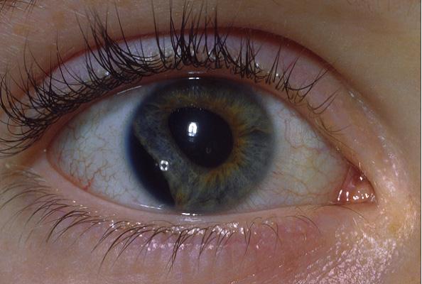
This 12 year-old boy was treated for a traumatic hyphaema after being hit with a stone. The above picture was taken two week later when he was reviewed in the clinic.a. What does the picture show?
Iridodialysis.b. What complications can develop in this condition?In iridodialysis, the base of the iris which is the thinnest and weakest area of the iris becomes separated from the ciliary body and the scleral spur. This type of injury is associated with a large hyphaema.
The complications resulting from iridodialysis include:Other associated complications include:
- Cosmesis. This is especially obvious in patients with light iris.
- Photophobia.
- Diplopia.
- Glaucoma. This can occur if there were significant angle recession. The incidence is increased if the recession involves more than 180 degrees of the angle.
- Hypotony. This can occur if there were associated cyclodialysis in which there is a tear between the ciliary body and the scleral spur.
c. How would you manage the patient?Treatment is not usually needed unless the patient has severe symptoms (cosmesis, photophobia and diplopia) due to the iridodialysis. In which case, the following options may be tried:
- painted contact lens
- surgical treatment in which the free iris root is sutured to the limbus.
| Click here for the questions | Click here for the main page | Click here for FRCOphth/MRCOphth
/FRCS tutorials |