 |
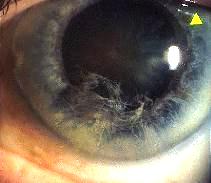
This 72 year old man was referred by his optician because of the appearance of his left eye as shown above. His visual acuity and intraocular pressures were normal in both eyes.a. What does the picture show and what is the diagnosis?
The picture shows ruptured inferior iris stroma with the anterior fibres floating free in the anterior chamber. It is typical of iridoschisis.Iridoschisis is an uncommon degenerative condition which occurs bilaterally in patients in their sixth and seventh decade. There is a split in the iris stroma separating it into the anterior and posterior parts. The posterior part remains attached to the dilator muscle and the retinal pigment epithelium whereas the anterior part is torn into fibres and floats freely in the anterior chamber.
b. What are the causes of this condition?Iridoschisis is usually seen as a degenerative process but may also occur in the following condition:
- iris necrosis from acute angle closure glaucoma
- iris trauma during cataract operation
- long-term use of miotics
- iris contusion
c. Is it necessary to follow-up this patient?Iridoschisis requires long-term follow-up as it is associated with glaucoma (both angle closure and open angle) in 50% of cases. The cause of glaucoma is unknown but it has been postulated that in the case of open angle glaucoma the cause may be due to pigment dispersion causing obstruction of the aqueous outflow. In the case of angle closure glaucoma, iridoschisis may be the result rather than the cause.In addition, the cornea may develop oedema due to endothelium damage from the free floating anterior fibres.
| Click here for the questions | Click here for the main page | Click here for FRCOphth/MRCOphth
/FRCS tutorials |