 |
Eyelids & the Anterior Segment:
Case twenty one
|
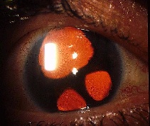
This is the anterior segment of a 40 year-old woman who was being treated for a right glaucoma. There was no history of trauma and the left eye was normal.a. What is the diagnosis?
Essential iris atrophy due to iridocorneal endothelial dystrophy (ICE). The pictures shows multiple iris holes (polycoria) and pupillary distortion (corectopia).b. How effective is anti-glaucoma treatment for this condition?ICE syndrome is typically unilateral and affects middle-aged women.ICE covers a spectrum of three conditions which have the shared abnormalities of iris, endothelium and the trabecular meshwork: Essential iris atrophy. Prominent polycoria and corectopia Chandler's syndrome. Mild iris atrophy and corectopia with hammered appearance of the corneal endothelium Cogan-Reese syndrome. Mild iris atrophy with pigmented nodules on the iris surface.
In ICE syndrome, the endothelium is abnormal and grows over the trabecular meshwork and the iris causing blockage of the trabecular meshwork.Initially the glaucoma can be controlled with medication but as the condition progresses, filtration operation become necessary. However, the endothelium can grow over the fistula and causes failed trabeculectomy.
c. What is the visual prognosis in this condition?The visual prognosis is usually poor because the glaucoma is difficult to control. In addition, the cornea often decompensates due to the abnormal endothelium and corneal graft may be required.
| Click here for the questions | Click here for the main page | Click here for FRCOphth/MRCOphth
/FRCS tutorials |