Eyelids & the Anterior Segment:
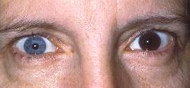
                   Case twenty seven


This 38 year-old woman had had a left intraocular foreign body removed one year ago. Her current visual acuity was 6/6 in the right eye and 6/60 in the left.

a. What is the most likely diagnosis ?

Ocular siderosis. 

The picture shows heterochromia with the left iris being darker. The dark iris resulted from the retention of  intraocular foreign body containing irion..


b. What other conditions can give rise to the above appearance?

Heterochromia iridis can be divided into those conditions which make the iris darker (hyperchromia) and those lighter (hypochromia). 

Hyperchromia 

  • idiopathic
  • iris naevus or melanoma
  • rubeosis iridis
  • siderosis
  • xanthochromia from hyphaema
  • unilateral use of latanoprost
Hypochromia 
  • idiopathic
  • congenital Horner's syndrome
  • trauma or surgery
  • previous inflammation such as Fuch's heterochormic uveitis


c. What would the electrophysiological tests reveal?

In early siderosis, the ERG shows increased a-wave with normal b-wave. As the condition progresses, the b-wave diminishes and in advanced cases the ERG is flat.


d. What other ocular signs may be present?

Siderosis damages the ocular structures mainly through the toxic effects of iron on the epithelium. In siderosis, the following signs may be seen: 
  • mydriasis due to damage of the sphincter muscle
  • glaucoma from damaged trabecular meshwork
  • cataract from iron deposition
  • pigmentary changes in the retina with attenuation of the vessels and optic atrophy
 
Click here for the questions Click here for the main page Click here for FRCOphth/MRCOphth
/FRCS tutorials