Figure 1 |
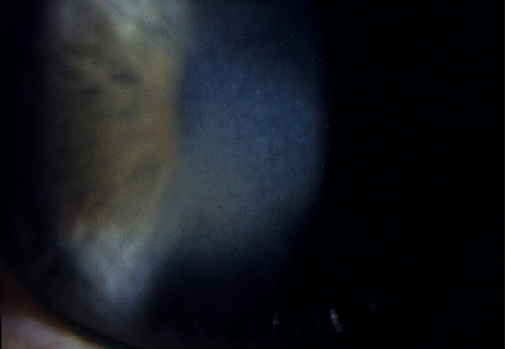
Figure 2 |
Eyelids & the Anterior Segment:
Case thirty one
|
Figure 1 |
Figure 2 |
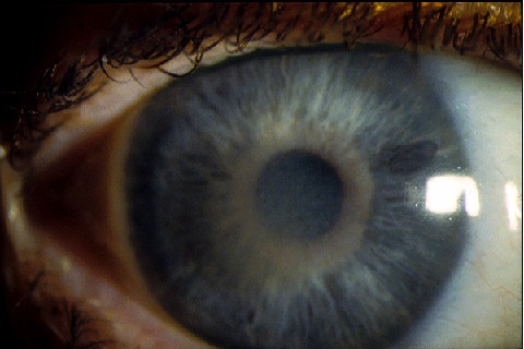
This 53 year-old woman was referred by his optician because of the above corneal appearance found in both eyes. Her corrected visual acuity was normal in both eyes.a. What do the pictures show?
Francois dystrophy (central cloudy dystrophy)Figure 1 show opacities which are denser centrally and fade peripherally. Figure 2 shows the central opacities to be in the deep stroma and appear as grey and polygonal areas without distinct margins. The opacities are separated by fine crack-like zones. (The appearance of the opacities have also been described as posterior crocodile shagreen).
b. Is this condition inherited?It is inherited in an autosomal dominant fashion. Therefore, examination of the family members is useful in confirming the diagnosis.c. What is the visual prognosis for this patient?
Francois dystrophy is a bilateral, symmetrical, non-progressive disorder. Most cases are discovered incidentally and the patients have no visual symptoms. No treatment is required. Both the corneal sensation and thickness are normal.
| Click here for the questions | Click here for the main page | Click here for FRCOphth/MRCOphth
/FRCS tutorials |