 |
Medical Retina & the Posterior Segment:
Case 21
|
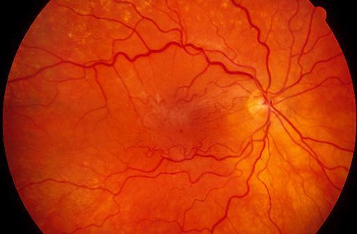
This 65 year-old woman complained of distorted vision in her right eye. Her visual acuity was 6/12 in the right eye and 6/6 in the left. Examination revealed the above right fundal appearance. a. What is the cause of her distorted vision?
Epiretinal membrane.The picture shows wrinkling and contraction of the macula with tortuosity of the small retinal vessels. The epiretinal membrane is made up mainly of glial cells and retinal pigment epithelial cells. The contraction of these cells along the internal limiting membrane is thought to be responsible for the signs and symptoms.
b. What conditions may give rise to this appearance?Conditions that can give rise to epiretinal membrane include:
- Idiopathic. In which no pathology is found.
- Retinal vascular disorders:
- proliferative retinopathies such as diabetes mellitus
- exudative retinovascular disorders such as diabetic maculopathy
- retinal vein occlusion
- vitreous haemorrhage- retinal tears or hole or detachment.
- ocular inflammatory disorders
- trauma
- after surgery such as cryopexy, photocoagulation and cataract operation
- congenital conditions such as retinitis pigmentosa
c. How would you manage this patient?It is important to examine for possible associated conditions especially retinal holes or tear which may require treatment.
Treatment is dependent on how disabling is the patient affected by the distortion. If the distortion is mild or non-troublesome then it is reasonable to review the patient at regular interval. However, in those much troubled by the distortion surgery is the only definitive treatment. This involves par plana vitrectomy and membrane peels.
| Click here for the questions | Click here for the main page | Click here for MRCOphth / FRCS tutorials |