Figure 1 |
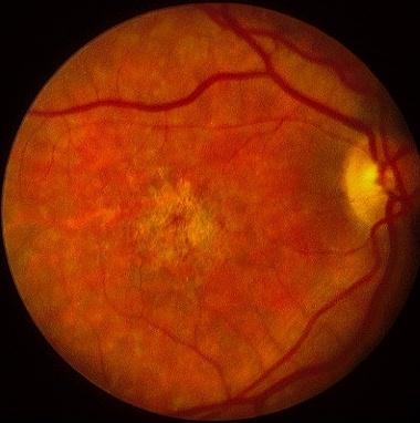
Figure 2 |
| Medical Retina & Posterior Segment: |
| Case 26 |
|
Figure 1 |
Figure 2 |
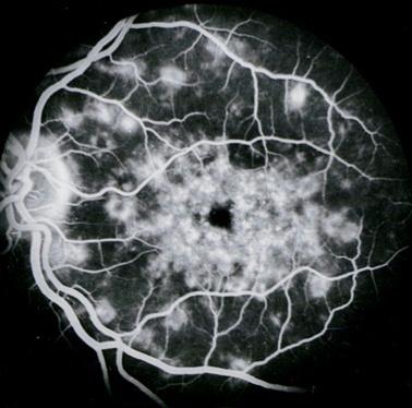
This 20 year-old man complained of a three year history of poor vision. His visual acuity was 6/12 in both eyes. The ERG was normal but the EOG was slightly reduced. The fluorescein angiography of the left eye is shown above. a. What is the most likely diagnosis?
Stargardt's disease.b. What is the histopathology of the lesions seen?The fluorescein angiography shows multiple subretinal hyperfluorescent lesions radiating from the macula. His right eye (figure 2) shows multiple yellowish flecks (or "fishtail" flecks) with atrophic area in the macula. The features and history are typical of Stargardt's disease.
Stargardt's disease is the most common form of juvenile macular dystrophy and is inherited mainly in an autosomal recessive fashion. The ERG is usually normal but the EOG subnormal due to involvement of the retinal pigment epithelium.
The lesions result from the accumulation of lipofuscin (acid mucopolysaccharide) within the retinal pigment epithelium.c. What is the visual prognosis of this condition?
This accumulation is often more extensive than observed on fundoscopy.The patient typically present in the second decade with slowly progressive decreased central vision. The visual acuity usually declined to 6/60 in the third or fourth decade due to macular atrophy.Diagnosis of Stargardt's disease may be difficult in the early stage before the appearance of the macular changes or the typical "fishtail" flecks. Fluorescein angiography may be useful at this stage as it may show "dark choroid" in the early phase. This is due to the accumulation of the lipofuscin within the RPE cells blocking the transmission of choroidal fluorescein.
Click here for the questions Click here for the main page Click here for MRCOphth / FRCS tutorials