 |
Medical Retina & the Posterior Segment:
Case seventeen
|
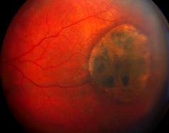
This 40 year-old man was referred by his optician because of the above lesion in his left peripheral fundus. He was otherwise fit and well and the visual acuity was 6/6 in both eyes.a. What is the most likely diagnosis?
Congenital hypertrophy of the retinal pigment epithelium.
b. What features suggest that this is not a choroidal naevus?Congenital hypertrohy of retinal pigment epithelium (CHRPE) is black and has a sharply demarcated border. The centre may be depigmented and the underlying choroid may be visible. The lesion has no malignant potential.In contrast, choroidal naevus is grey in colour with a ill-defined border. Drusen may be present on its surface. A small proportion of choroidal naevus may develop into melanoma.
c. What is the significance of this lesion?CHRPE is usually found incidentally and have no clinical significance. However, the presence of multiple CHRPE is associated with Garner's syndrome ( a autosomal dominant condition associated with familial adenomatous polyposis of the colon and mesodermal tumours such as dermoid tumours and osteoma of the skull).
| Click here for the questions | Click here for the main page | Click here for FRCOphth/MRCOphth
/FRCS tutorials |