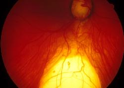
| This is the right fundal appearance of a 24 year-old man who has
a normal visual acuity.
a. What is the diagnosis?
Coloboma of the retina and choroid.
The picture shows a defect in the inferior fundus allowing the white
sclera to become visible.Visual field will shows a superior field defect.
b. What is responsible for this appearance?
It is caused by the incomplete closure of the embryonic fissure
of the optic cup (the closure is usually completed in the fifth week of
gestation). As the fusion occurs inferonasally, a typical coloboma is seen
inferonasally.The coloboma can also affects the iris and optic nerve.
Provided the coloboma does not involve the macula the vision is usually
normal.
c. What complications may occur with this condition?
The following may be associated with coloboma:
-
CHARGE syndrome (Coloboma, Heart defects, Atresia of choanae, Retarded
growth and development, Genital hypoplasia and Ear anomalies including
deafness)
-
increased incidence of rheumatogenous retinal detachment. A break in the
thinned retina (intercalary membrane) overlying the defect is often responsive
for this.
|