Figure 1 |
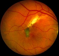
Figure 2 |
Medical Retina & the Posterior Segment:
Case 24
|
Figure 1 |
Figure 2 |
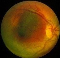
This 32 year-old painter was accidentally hit in the right eye from an unhinged cabinet door. On arrival in the casualty, his visual acuity was counting finger in the right eye. Slit-lamp examination revealed a right hyphaema and the above appearance in Figure 1. Two months after the injury, his vision returned to 6/9 and the appearance in Figure 2 is noted. a. What is the diagnosis?
Choroidal rupture.Figure 1 shows the presence of blood in the subretinal and the sub-RPE region (blood in the sub-RPE appears darker than those in the subretinal due to masking by the RPE). Figure 2 shows choroidal rupture (white crescenteric lesion) with pigmentary changes in the macular area.
b. What is the mechanism of the injury?In blunt trauma, the eye wall is stretched. Of the three layers of structures, the sclera is strong and the retina is elastic but the Bruch's membrane that forms the layer between the RPE and choriocapillaries is neither strong nor elastic. As a result, it is susceptible to rupture. When this occurs, the Bruch's membrane retract taking within the overlying RPE and the inner layers of the choroid making the white sclera visible. Subretinal haemorrhage is a common associated sign.
c. Is he at risk of further visual loss from the above appearance?The presence of choroidal rupture increases the risk for the development ofchoroidal neovascularization within the break in Bruch's membrane. This may occur months or years after the injury. Therefore, the patient should be followed regularly and advised to seek help if there were any development of distorted vision. Laser photocoagulation is useful in treating such complication.
| Click here for the questions | Click here for the main page | Click here for MRCOphth / FRCS tutorials |